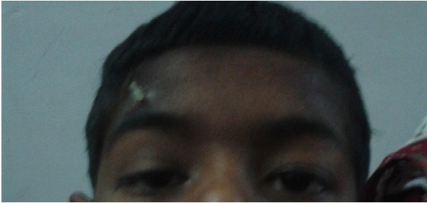
Marcus-Gunn Jaw Winking Ptosis
All content on Eyewiki is protected by copyright law and the Terms of Service. This content may not be reproduced, copied, or put into any artificial intelligence program, including large language and generative AI models, without permission from the Academy.
Marcus Gunn jaw-winking first was described by Robert Marcus Gunn in 1883 in a 15 year old girl as unilateral blepharoptosis with associated upper eyelid contraction. It is the most common form of congenital neurogenic ptosis. [1]
Disease Entity
Disease
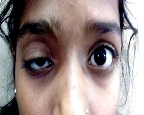
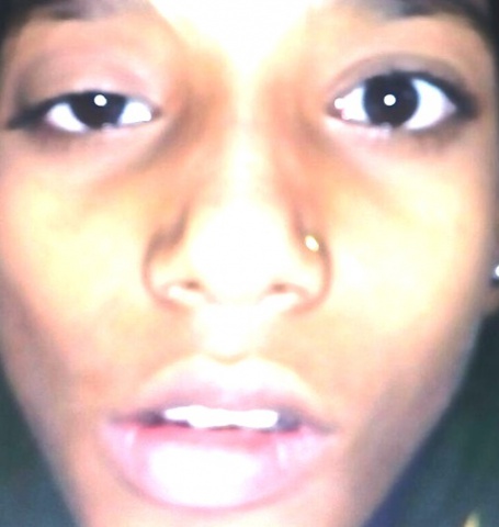
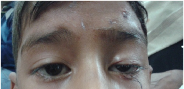
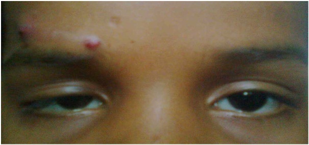
Marcus Gunn jaw-winking ptosis is a congenital ptosis associated with synkinetic movements of the upper lid on masticating movements of the jaw. It is usually unilateral but rarely presents bilaterally. It affects males and females in equal proportion. [2]
Etiology
This synkinetic movement results from a congenital aberrant connection between motor branches of the trigeminal nerve controlling muscles of mastication and superior division of the oculomotor nerve controlling the levator palpebrae superioris. Although most cases of Marcus Gunn phenomena (MGP) are said to be congenital, acquired forms of this condition are also known to exist.[3] Marcus Gunn phenomena may develop after eye surgery, syphilis, trauma, pontine tumors, etc. Spontaneous remission of the acquired form may occur, whereas the congenital form typically persists with little to no improvement over life.
General Pathology
Marcus Gunn jaw-winking ptosis is believed by most to be due to abnormal innervation of the levator muscle and not secondary to myopathic changes, so most histopathologic studies have revealed normal striated muscle. Some studies have found variable degrees of fibrosis within the affected levator muscle. [4]
Pathophysiology
Electromyographic studies demonstrate this synkinetic innervation by showing simultaneous contraction of the external pterygoid and levator palpebrae superioris muscle. In rare cases, synkinesis may be present between the internal pterygoid and levator muscles. In these cases, the eyelid elevates on closing the mouth and clenching the teeth.
A complete explanation has not yet been derived to elucidate the rationale of jaw-winking phenomenon.
Various theories have been hypothesized.
Aberrant connection
This hypothesis is favored by many. The location of the aberration may differ as follows :
- Cortical or sub cortical connections
- Internuclear connections or faulty distribution in the posterior longitudinal bundle
- Infranuclear connection exists between motor branches of the trigeminal nerve (CN V3) innervating the external pterygoid and the fibers of superior division of the oculomotor nerve (CN III) that innervates the levator muscle of the upper lid
- Peripherally - some CN V fibers may reach the levator via the auriculo-temporal nerve.
Functional Interference
- Irritation of normally dormant connection
- Disinhibition of pre-existing phylogenetically more primitive mechanisms (Ascher): This is thought to explain why individuals who are not affected will often open their mouth while attempting to widely open their eyes to place eye drops
- Spread of impulses by irradiation
Atavistic Reversion
- Atavism meaning evolutionary throwback, just as in fish a strong associated movement of jaw opening and eye opening i.e., deep muscle contracting and superficial muscle relaxing. Similarly a weak levator palpebrae superioris may only elevate the lid when its antagonist, the orbicularis (superficial muscle) is reflexively relaxed by jaw opening (external pterygoid-deep muscle contraction).
- EMG study suggested dysfunction in the midbrain and brainstem
Diagnosis
History
This phenomenon is typically first observed by the mother when the child is breastfeeding or bottle feeding. The history is typically of elevation of the ptotic lid then the child feeds. Past history of any ocular trauma or strabismus surgery may also be present.
Signs
- Blepharoptosis, usually unilateral
- Upper eyelid movement is seen on :
- Mouth opening
- Jaw movement toward the contralateral side
- Chewing
- Sucking
- Jaw protrusion
- Clenching teeth together
- Swallowing
- Cover Test : Hypotropia on involved side may be present due to associated superior rectus palsy.
Symptoms
Most common presenting feature is ptosis of the upper lid, which is associated with movement of upper eyelid noticed by the parents when the child eats or in case of an infant, while breastfeeding.
For more images see link https://www.aao.org/education/image/marcus-gunn-jaw-winking-syndrome-2
Clinical diagnosis
The diagnosis is made conclusively based on the signs as described above.
Diagnostic procedures
The amount of jaw-winking is the excursion of the upper eyelid with synkinetic mouth movement.
It is measured with a millimeter ruler.
Jaw-winking is assessed as:
- Mild < 2mm
- Moderate 2-5mm
- Severe ≥ 6mm
Additional testing
Abnormal oculocardiac reflex has been seen in patients with Marcus Gunn jaw-winking syndrome, so preoperative electrocardiogram (EKG or ECG) may be considered prior to surgery and the anesthesia team may be alerted to this unique feature of this syndrome.[5]
Associations
Ocular
- Strabismus (50%-60%)
- Superior Rectus Palsy-25%
- Double Elevator Palsy-25%
- Anisometropia - (5%-25%). Incidence of anisometropia among patients with Marcus Gunn jaw-winking syndrome is reported to be 5-25%.
- Amblyopia - (30-60%). Almost always secondary to strabismus or anisometropia, and rarely, is due to occlusion by a ptotic eyelid.
Systemic
Systemic anomalies in association with Marcus Gunn phenomenon are rare:
- Cleft lip/ Cleft palate
- CHARGE Syndrome reported in association with bilateral cases.
- Renal calculi
Other synkinetic abnormalities which may be associated with eyelid ptosis are Inverted Marcus Gunn Phenomenon and Marin-Amat syndrome. Initially, they were believed to be the same disease, but recent studies have found differences between them. In inverse Marcus Gunn phenomenon the upper lid falls to cover the eye with movements of mastication. This is more commonly seen as an acquired rather than congenital anomaly in central nervous system (CNS) disease. Marin Amat syndrome would have more of a blepharospasm like closure secondary to action of orbicularis oculi. The synkinesis is caused by aberrant regeneration of the seventh nerve with sprouts of axons supplying more than one muscle group. It represents patients with a connection between the orbicularis oculi and not with levator palpebrae superioris.
Management
Medical therapy
If amblyopia is encountered, treat aggressively with occlusion therapy and/or correction of anisometropia prior to any consideration of ptosis surgery.
Medical follow up
Six monthly follow up is usually advocated. Photographs can be helpful in monitoring patients. The patient should be checked for astigmatism due to the compression by the ptotic eyelid.
Surgery
The jaw-wink is considered cosmetically significant if it is 2 mm or more. An attempt to repair only the ptosis without correcting the jaw winking would result in an exaggeration of the aberrant eyelid movement to a level above the superior corneal limbus, which would be cosmetically unacceptable to the patient.
Dillman and Anderson stated that removal of a portion of the levator muscle above the Whitnall’s ligament (i.e., myectomy) is adequate to obliterate its function without extensive dissection and damage to eyelid structures. [6]
Beard and others have advocated bilateral excision of the levator muscle and bilateral frontalis suspension. This approach almost completely eliminates the wink and results in better symmetry, it is often difficult to persuade the parents and the patient to perform surgery on the contralateral normal eye.
Kersten et al advocate unilateral levator muscle excision and frontalis sling only on the affected side. [7]
If the postoperative result is judged to be unsatisfactory, the parents or the patient can opt for further surgery to the contralateral side. Any amblyopia and strabismus should first be addressed, as there may be insufficient drive to lift the disinserted eyelid. Relative contraindications are poor Bells' phenomena, reduced corneal sensations or dry eye can produce exposure keratopathy. Thus if operated, close follow up is needed.
Surgical follow up
Surgical follow up is necessary every 2-4 weeks to look for complications as described below.
Complications
Complications following ptosis surgery include:
- Undercorrection [8]
- Overcorrection
- Lagophthalmos
- Suture granuloma
- Sling slippage
- Sling extrusion
- Asymmetric lid crease
- Exposure keratitis
Amblyopia occurs in 30-60% cases
Prognosis
Prognosis is usually good in majority of cases. With careful observation and treatment, amblyopia can be treated successfully.
References
- ↑ Gunn RM. Congenital ptosis with peculiar associated movements of the affected lid. Trans Ophthal Soc UK. 1883; 3:283-7.
- ↑ Demirci H, Frueh BR, Nelson CC. Marcus Gunn Jaw-Winking Synkinesis Clinical Features and Management. Ophthalmology 2010;117:1447–1452.
- ↑ https://rarediseases.info.nih.gov/diseases/6972/marcus-gunn-phenomenon
- ↑ 4. Baldwin HC, Manners RM. Congenital blepharoptosis: a literature review of the histology of levator palpebrae superioris muscle. Ophthal Plast Reconstr Surg.2002;18:301-7.
- ↑ Pandey, M., Baduni, N., Jain, A., Sanwal, M. K., & Vajifdar, H. (2011). Abnormal oculocardiac reflex in two patients with Marcus Gunn syndrome. Journal of anaesthesiology, clinical pharmacology, 27(3), 398.
- ↑ Dillman DB, Anderson RL. Levator myomectomy in synkinetic ptosis. Arch Ophthalmol 1984; 102(3): 422-3.
- ↑ Kersten RC, Bernardini FP, Khouri L, et al. Unilateral frontalis sling for the surgical correction of unilateral poor-function ptosis. Ophthal Plast Reconstr Surg. 2005; 21(6):412-6; discussion 416-7.
- ↑ Park DH, Choi WS, Yoon SH. Treatment of jaw winking syndrome. Ann Plast Surg 2008; 60(4): 404-9.


