Iris Varix
All content on Eyewiki is protected by copyright law and the Terms of Service. This content may not be reproduced, copied, or put into any artificial intelligence program, including large language and generative AI models, without permission from the Academy.
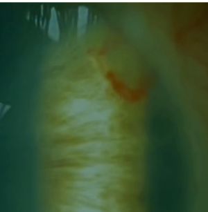
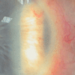
Iris varix is a condition characterized by elongated and dilated veins within the iris of the eye. It can be mistaken for other conditions such as iris hemangioma or melanoma. It is typically unilateral, often located in the inferotemporal quadrant of the iris, and can have a radial or circumferential orientation. It may be associated with features like a dilated episcleral sentinel blood vessel but does not exhibit characteristics of malignancy. Iris varix cases remains stable during follow-up, and no secondary tumors were observed.
Disease Entity
Disease
Iris varix is an elongated, dilated, typically tortuous vein. Iris varices predominantly unilateral varices (92.3%), with a minority being bilateral (7.7%). Single varices are more common (64.3%) than multiple varices (35.7%), with circumferential and bilateral varices exclusively being rare. Iris varices were frequently located in the inferotemporal (75%) and superotemporal (39.2%) quadrants, with some eyes having involvement in more than one quadrant (17.9%). Circumferential varices were limited to the inferotemporal quadrant. Radial orientation was most common (57.1%), followed by circumferential (21.4%), and some eyes exhibited both radial and circumferential varices.[1]
Etiology
Unknown.
Risk Factors
Unknown.
General Pathology
Unknown.
Pathophysiology
Unknown.
Diagnosis
Diagnosis is made clinically. But Fluorescein angiography (FA), UBM, or OCT-angiography can be helpful to distinguish varicose from other entities.
History
These lesions do not progress and could be congenital in nature.
Physical Examination
Signs
An iris vein that is elongated, enlarged, and typically tortuous, either radially or circumferentially. This can be differentiated from neovascular changes, which are typically much finer vessels and are usually not focal/sectoral.
Symptoms
Most of the cases are asymptomatic.
Clinical diagnosis
Diagnostic procedures
- Slit-lamp photography: utilizing side-by-side comparisons over time to monitor changes in anterior segment anomalies.
- Gonioscopy: focused on angle characteristics, such as open, narrow, or focal angle closure, pigment dispersion, and bridging blood vessels.
- High-frequency ultrasound imaging: involves recording longitudinal and transverse images in the central meridian of the varix, followed by a 360° screening of the entire anterior segment. Ultrasonography assesses iridociliary angle blunting, maximum iris thickness (meridian of the varix), adjacent ciliary body thickness (meridian of the varix), and iris pigment epithelium (IPE) characteristics (intact, erosion, posterior bowing, or anterior displacement). Iris nodularity and vascular channels can also be measured, with iris thickness exceeding 0.7 mm considered increased based on normative values.
- Fluorescein angiography (FA) of anterior segmen: FA is not a routine procedure but, when performed, may show hyperfluorescence appearance without dye leakage. Trombosed iris varices may show no perfusion in the area of mass and a regular perfusion in iridal network.
Laboratory test
No laboratory test is required. Pathological examinations reveal dilated capillaries with CD31 immunoperoxidase stain marked endothelial cells, without a proliferative pattern, consistent with a varix.[2]
Morphological categorization
Different orientations of iris varix were defined based on clinical visibility.
- Radial iris varix was identified when a visible portion traversed from the root of the iris to the pupillary margin in one or more meridians.
- Circumferential iris varix was characterized by a clinically visible portion running parallel to the pupillary margin.
- Combined iris varix included both radial and circumferential components.
Differential diagnosis
- Iris hemangioma
- Iris melanoma
- Iris AVM
- Diffuse neonatal hemangiomatosis (DNH)
- Hereditary Hemorrhagic Telangiectasia / Osler-Weber-Rendu Syndrome
- Wyburn-Mason Syndrome
- Iris Neovascularization
Management
Observation.
General treatment
No treatment is recommended in uncomplicated cases. Recurrent hyphema may need surgical intervention.
Medical therapy
No medical therapy is available.
Medical follow up
There is no recomendation on how long or how often they should be followed up.
Surgery
Resection may be considered in the event of repeated bleeding to mitigate the potential risk of secondary glaucoma [3]. Laser photocoagulation theoretically can be employed also. If there are concerns about a potential melanoma, it is advisable to conduct a thorough examination and pathological assessment [4].
Surgical follow up
No recurrence reported in the available literature.[1]
Complications
Iris varices experiencing repeated hemorrhages into the anterior chamber may result in secondary glaucoma.[3] When these veins become thrombosed they may resemble iris melanoma.[4]
Prognosis
Over a 10-year follow-up period, stability was observed in 96.4% of eyes, with only one eye (3.6%) showing regression. There were no occurrences of corectopia, ectropion uveae, hyphema, or metachronous anterior segment benign or malignant tumors. [1]
References
- ↑ 1.0 1.1 1.2 Jain P, Finger PT. Iris varix: 10-year experience with 28 eyes. Indian J Ophthalmol. 2019 Mar;67(3):350-357. doi: 10.4103/ijo.IJO_1253_18. PMID: 30777952; PMCID: PMC6407407.
- ↑ Broaddus E, Lystad LD, Schonfield L, Singh AD. Iris varix: report of a case and review of iris vascular anomalies. Surv Ophthalmol. 2009 Jan-Feb;54(1):118-27. doi: 10.1016/j.survophthal.2008.10.005. PMID: 19171213.
- ↑ 3.0 3.1 Ang LP, Sim DH, Chiang GS, Yong VS. Iris varix. Eye (Lond). 1997;11 ( Pt 5):733-5. doi: 10.1038/eye.1997.187. PMID: 9474328.
- ↑ 4.0 4.1 Shields JA, Shields CL, Pulido J, Eagle RC Jr, Nothnagel AF. Iris varix simulating an iris melanoma. Arch Ophthalmol. 2000 May;118(5):707-10


