Intravitreal Injection Assistive Devices
All content on Eyewiki is protected by copyright law and the Terms of Service. This content may not be reproduced, copied, or put into any artificial intelligence program, including large language and generative AI models, without permission from the Academy.
Intravitreal injections are among some of the most common procedures in ophthalmology. Among the indications include diabetic retinopathy, neovascular age-related macular degeneration, retinal vein occlusion with macular edema, and choroidal neovascularization from various etiologies.
In response to an increasing demand, some physicians have begun using intravitreal injection assistive devices to help deliver faster, safe, and accurate injections.
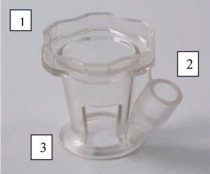
InVitria Injection Assistant (FCI Ophthalmics, Pembroke, MA)

The InVitria is a single-use, clear, conjunctiva-fixated mould developed in 2013 and distributed by FCI Ophthalmics. It is made of polycarbonate and is composed of a guide tube designed to deliver injections 3.50 mm beyond the limbus. The guide tube is angulated at 28 degrees and provides a 5.60 mm fixed depth insertion of a 30-gauge needle.
Available Studies
A study by Ratnarajan et al used the InVitria in a series of injections without the use of a lid speculum, caliper and surgical drapes. They reported an improvement in pain reduction, globe stability, needle entry reproducibility and cost-effectivity compared with the conventional technique. [2] This device has also been shown to aid in the successful training of ophthalmic nurses in delivering intravitreal injections in the United Kingdom.[3] Baneke et al, however, advises against its use in post-trabeculectomy patients as its mould-like structure poses a risk for bleb-related complications.[4]
Rapid Access Vitreal Injection (RAVI) Guide (Katalyst Surgical, Chesterfield, MO)

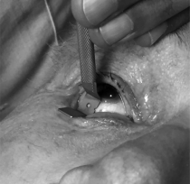
The RAVI guide is an intravitreal assistive device that is composed of a square baseplate and a handle. The baseplate measures 7 mm x 7 mm and has an approximately 1.5-mm-diameter hole in the center and flanges on opposite sides. The handle is attached to one of the flanges. The location and size of the hole allows for injection within a distance of approximately 3 mm to 4 mm from and 360˚ around the limbus. The device is autoclavable and reusable, and it can replace the function of both the lid speculum and caliper.
Available Studies
The device has been shown to be associated with similar pain scores as those of the traditional technique, while successfully separating the eyelids, and guiding injection of the needle.[6]
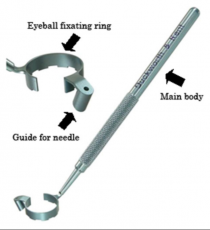
Doi-Umeatsu Intravitreal Injection Guide (Duckworth & Kent Ltd., England)

The Doi-Umeatsu Intravitreal Injection guide is a lightweight, titanium instrument composed of three parts: the main body, fixation ring and the needle guide. The main body is an elongated rod which primarily serves as the handle. It is connected at an obtuse angle to an incomplete (approximately 315˚) circular ring for fixation of the globe. The ring is ridged to facilitate good contact and control of the patient’s eye. The needle guide has a 0.5 mm hollow lumen to allow the injection of a needle at a guarded depth of approximately 8 mm.
Available Studies
A study by Watanabe et al has demonstrated that the use of the guide in intravitreal injections is safe, even for physicians with limited experience.[8]
Malosa Intravitreal Injection Guide (Beaver-Visitec International, Waltham, MA)

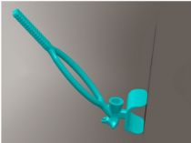
The Malosa Intravitreal Injection Guide is a single-use, sterile, polycarbonate instrument developed in the United Kingdom by Dr. Salman Waqar. It is composed of a lash guard, a triangular footplate, a needle chamber and a handle. An apex indicator and fixation studs are found in the footplate for accurate limbal placement and globe stabilization, respectively. The cylindrical chamber guides precise and controlled needle entry at a fixed depth. The wishbone shape of the handle aids in adequate visualization during the procedure.
Available Studies
Published studies have reported good patient feedback and minimal reduction in intraprocedural pain in comparison with the traditional technique using a lid speculum.[10][11]
Robot-Assisted Injection Systems
Early 2025 saw preclinical progress in robotic-assisted intravitreal injection systems, building on prior proposals (e.g., Weiss et al., 2018), with a focus on automation for precision and tremor reduction.
A January 2025 paper reported on a prototype robotic arm (University of California, Irvine) tested in porcine eyes. The device uses real-time OCT imaging and a 30-gauge needle guided by a 6-axis manipulator, achieving a targeting accuracy of ±50 µm (vs. ~200 µm freehand). In 20 trials, it reduced injection time by 22% (mean 45s vs. 58s) and eliminated hand tremor, a factor in 5–10% of freehand complications like vitreous hemorrhage. Human trials are pending, but the system integrates with standard anti-VEGF vials (e.g., aflibercept), suggesting compatibility with existing workflows.[12]
Robotic assistance could standardize injections, reduce operator fatigue, and lower rare but severe complications (e.g., retinal detachment, 0.01–0.05% risk). Cost and scalability remain barriers, with no commercial timeline yet.
Automated Injection Guide
An automated assistive system for intravitreal injection has been proposed and was demonstrated to be effective in delivering precise injections in porcine eyes.[13] No known human studies have been reported.
Additional Resources
- Video: Invitria - Intravitreal Injection Device by FCI. https://www.youtube.com/watch?v=w14uh44RSRk
- Video: Doi-Umeatsu Intravitreal Injection Guide. https://www.youtube.com/watch?v=U-1hYkSIynU&t=128s
- Video: Malosa Intravitreal Injection Guide. https://www.youtube.com/watch?v=USc80RIhOkA
References
- ↑ Ratnarajan G, Nath R, Appaswamy S, Watson SL. Intravitreal injections using a novel conjunctival mould: a comparison with a conventional technique. Br J Ophthalmol. 2013;97(4):395–397.
- ↑ Ratnarajan G, Nath R, Appaswamy S, Watson SL. Intravitreal injections using a novel conjunctival mould: a comparison with a conventional technique. Br J Ophthalmol. 2013;97(4):395–397.
- ↑ Michelotti MM, Abugreen S, Kelly SP, et al. Transformational change: nurses substituting for ophthalmologists for intravitreal injections - a quality-improvement report. Clin Ophthalmol. 2014;8:755–761. Published 2014 Apr 15.
- ↑ Baneke AJ, Vakros G, Sharma V, Wong SC. BLEB-RELATED ENDOPHTHALMITIS AFTER USE OF THE INVITRIA INJECTION GUIDE. Retin Cases Brief Rep. 2020;14(1):33–34.
- ↑ Han DP, McKenney KC, Kim JE, Weinberg DV, Musch DC, Singh RS. CLINICAL EVALUATION OF THE RAPID ACCESS VITREAL INJECTION GUIDE: A Handheld Instrument for Assisting Intravitreal Injections. Retina. 2017;37(4):778–781.
- ↑ Han DP, McKenney KC, Kim JE, Weinberg DV, Musch DC, Singh RS. CLINICAL EVALUATION OF THE RAPID ACCESS VITREAL INJECTION GUIDE: A Handheld Instrument for Assisting Intravitreal Injections. Retina. 2017;37(4):778–781.
- ↑ Watanabe K, Masafumi U, Mohamed YH, Watanabe T, Doi Y, Azusa F, Kitaoka T. Safety of Intravitreal Injection Guide. J Vitreoretin Dis. 2017;2(1):26–31.
- ↑ Watanabe K, Masafumi U, Mohamed YH, Watanabe T, Doi Y, Azusa F, Kitaoka T. Safety of Intravitreal Injection Guide. J Vitreoretin Dis. 2017;2(1):26–31.
- ↑ Waqar S, Ayaz A, Karamat I, et al. A novel device for rapid, safe and precise delivery of intravitreal injections. BMJ Innov Epub ahead of print: [22 March 2020]. doi:10.1136/ bmjinnov-2018-000337
- ↑ Waqar S. Comment on: Optimisation of intravitreal injection technique using a Barraquer speculum with solid flat blade and finger stabilisation. Eye (Lond). 2019;33(3):512.
- ↑ Waqar S, Ayaz A, Karamat I, et al. A novel device for rapid, safe and precise delivery of intravitreal injections. BMJ Innov Epub ahead of print: [22 March 2020]. doi:10.1136/ bmjinnov-2018-000337
- ↑ Patel, R., et al. (2025). Robotic-assisted intravitreal injection: Preclinical evaluation in porcine models. Investigative Ophthalmology & Visual Science, 66(1), 89–97. DOI: 10.1167/iovs.66.1.89.
- ↑ Ullrich F, Michels S, Lehmann D, Pieters RS, Becker M, Nelson BJ. Assistive Device for Efficient Intravitreal Injections. Ophthalmic Surg Lasers Imaging Retina. 2016;47(8):752–762.

