History of Ophthalmology
All content on Eyewiki is protected by copyright law and the Terms of Service. This content may not be reproduced, copied, or put into any artificial intelligence program, including large language and generative AI models, without permission from the Academy.
“Those about to study medicine, and the younger physicians, should light their torches at the fires of the ancients” – Baron Carl von Rokitansky (1804-1878)
Ophthalmology has always been at the forefront of medical research while having an admirable history of innovation. As we continue to embrace the rapid advances in various development and technologies in ophthalmology, we would do well to take a step back and appreciate its incredible journey as a specialty.
Prehistoric & Classical Era (3000 BC to AD 476)
Ancient Egypt
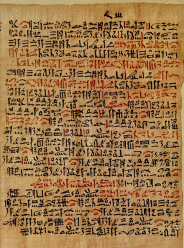
The practice of ophthalmology has been documented since ancient times. Its existence can be traced back to Ancient Babylon with a reference to the eyes made in the Code of Hammurabi (2250 BC) – “If a physician performs eye surgery and saves the eye, he shall receive ten shekels in money” [1]. However, it was not until Ancient Egypt where the first known written record of medical treatments of eye disease was discovered in the Ebers Papyrus (Fig. 1), a 110-page scroll that dates back to 1550 BC [2].
The Ebers Papyrus paid significant attention to the eyes, with nine pages of this ancient medical manuscript devoted to eye conditions. Various conditions such as pterygium, staphyloma, trichiasis, cataract and ophthalmoplegia were described in detail with magical formulas and folk remedies ranging from tortoise brain to cow’s blood for such afflictions [3].
The eye also played a significant role in ancient Egypt and featured heavily in everyday secular and religious activities. A famous example of this is the Eye of Horus (Fig. 2), which was ripped out by Seth and later restored magically by Thoth, after which it was called wedjat ('The Whole One') [4]. It became one of the most popular amulets worn by the Egyptians and featured as a prominent symbol of healing, protection and good health.
Ancient Greek
Pre-Hippocratic era
Next came the pre-Hippocratic era, where the anatomical conception of the eye was mainly speculative. Alcmaeon of Croton (540-500 BC), a prominent philosopher-physician at the time, was widely regarded as the first to examine and describe the human eye. He and others hypothesized that the eye to comprise two layers: 1) an outer layer consisting of the sclera and cornea and 2) an inner layer bordered anteriorly by the pupil with fluid at the center [5]. He also described the eye to be attached to the brain by ducts (poroi) through which a special fluid, believed to the medium of vision, flowed from the eye to the brain.
Hippocratic era
The Hippocratic Corpus, a collection of approximately 60 early Ancient Greek medical works by Hippocrates (460-375 BC), further advanced the ideas of Alcmaeon of Croton [6]. The eye was now described as comprising three layers (not two): 1) a thick outer layer, 2) a thinner middle layer which may protrude like a bladder when injured and 3) an inner layer which is thinnest and very prone to damage. Aristotle (384-322 BC) further put forth the existence of this anatomical framework based on animal dissections and described the optic nerve as a duct connecting the eye with the membranes of the brain (Fig. 3) [7].
Significant emphasis was also placed on the prognostic value of the eyes in the context of clinical medicine. Notable excerpts from the Hippocratic Corpus highlighting these include [8]:
- The eyes normally sparkle in tuberculosis (Epidemics iii. 14)
- Squinting eyes in postpartum fever is prognostically bad (Epidemics iii. 11)
- Rapid eye movement in the presence of epigastric pathology reflects insanity (Prognostic vii)
- The prognosis is bad when the eyes are distorted in fever or become blind (Aphorisms iv. 45)
Hellenistic (Post-Hippocratic) era
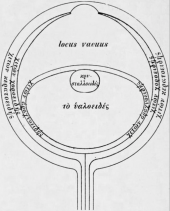
The invasion of the Achaemenid Empire in 330 BC by Alexander the Great ushered in the Hellenistic era in which Greek cultural influence and power reached the peak of its geographical expansion. The Alexandria School of Medicine, where the first systematic dissection of the human body was performed, further advanced the understanding of ocular anatomy and physiology [10]. Much of this was contributed by Herophilus (330-260 BC), a Greek physician who showed a special interest in the eye and devoted a treatise specifically to this topic [11]. Unfortunately, only fragments of his written works survived, with much of it inherited through quotations from subsequent scholars such as Celsus, Rufus and Galen. Through their work, existence of further ocular structures was given clear recognition, including the lens (a drop-like body named crystalloides) and a large empty space (locus vacuus) containing “humor”, likely representing the anterior chamber (Fig. 4).
Ancient India
Although the knowledge and history of ophthalmology in this region has been largely insular for hundreds of years, it is seeing a rapid resurgence as a result of recent restoration initiatives. It is clear that ophthalmology has been practised from the earliest times but the interpretation of certain findings has remained the subject of debate.
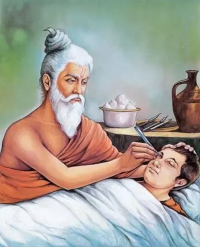
Sushruta, the most celebrated physician and surgeon in India, is widely regarded as the father of Indian ophthalmology. The exact timeline of his existence remains a controversial topic due to a lack of traceable evidence but current consensus places him around the fifth century BC (a century earlier than Hippocrates) [12]. He devoted a complete volume of his experiences on ocular anatomy and pathology to the final sections of the Sushruta Samhita, a monumental treatise on surgery. This compilation, referred to as the Uttara Tantra, contained elaborate descriptions of 76 eye diseases (Nayana-Budbada), of which 51 were amenable to surgical treatment. These included diseases such as glaucoma (Gambhirika), uveitis (Adhi-mantha), cataract (Kacha) and trichiasis (Pakshma-kopa) [13]. In addition, Sushruta described in the Samhita of what may be the first extracapsular cataract extraction using the Yava Vaktra Salaka, a sharply curved needle used to loosen and push the cataract out of the field of vision (Fig. 5) [14].
Ancient Rome
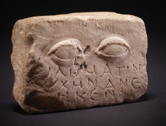
Roman ophthalmology in classical antiquity represented further progress and modification of pre-existing Greek knowledge. This was reflected by the fact that most physicians working in the Roman Republic were in fact from the Hellenistic area and worked as slaves in wealthy families. Pliny the Elder, a commander of the early Roman empire, commented on this in his encyclopedic Naturalis Historia which claimed that Rome had well-established medical care but no Roman physicians [15]. Trachoma (called “aspiritudo” (roughness)) was particularly endemic in ancient Rome and was treated with collyria containing copper acetate and surgical removal of the granular lesions [16]. Various sculptures were dedicated to the eyes, evident through existing stone votives for ocular ailments left in shrines by grateful patients (Fig 6.).
The Greco-Roman physicians made remarkable anatomical discoveries of the eye and refined surgical instruments and techniques. Among these, Celsus (25 BC to 50 AD) was the first to systematically deal with ophthalmology, with related manuscripts preserved to the present day. Through his pioneering work De Re Medica, Celsus earned the reputation as one of the greatest contributors to ophthalmology during the Roman empire [17]. In it, he described in Latin various ocular diseases and their treatment. These descriptions, largely inspired by his Greek predecessors, were later developed into a more sophisticated classification by the Romans. Certain views such as the crystalline lens as the central organ of vision and cataract as opaque concretions between the lens and iris were still held with high regard, and these persisted through the Middle Ages [18].
The Middle Ages (AD 476 to 1450)
Arabian Era
During the Middle ages or medieval period, the fall of the Western Roman Empire saw the centre of Caucasian civilization shifting eastward to the Arabist world which dominated not only politically but intellectually and culturally. The rich heritage of the Grecian era was largely neglected by Europe during the dark period, in contrast to the Arab Empire which embraced and further developed Hellenistic medical knowledge. During this period, incomparably more manuscripts on ophthalmology were written in Arabic compared to Latin, Greek and other European languages combined [19].
The Kahhal (کحال) or oculist, was highly regarded in society and had a privileged place in royal households. The most famous Kahhal, not only during his time but cited as an authority throughout the medieval period, was Hunayn ibn Ishaq al-Ibadi (also known as Johannitius in Latin; 809-873). He was the son of a Nestorian Christian chemist with knowledge of the Greek language and wrote the Book of the Ten Treatises of the Eye which built upon the work of Greek scholars and delineated the first detailed anatomical illustration of the eye [20].
Revolutionary work in the field of optics was achieved by Ibn Al-Haytham (also known as Alhazen in Latin; 965-1040) through his Book of Optics (Kitab al-Manazir). In it, he described the correct theory of vision in which the seeing process was facilitated by light rays radiating from an object to the eye (Fig. 7) [21]. This disproved the opposite ideas of preceding Greek philosophers who proposed the intromission theory, whereby rays emanated from the eye itself to an object.
The previous practices of earlier Greco-Roman physicians were deemed crude and frowned upon by many of the leading figures in the medieval Arabist age. Guided by scientific methods, the flourishing science of ophthalmology in the Arabic era served as the foundation to that of European science and endured well into the Modern era. This was echoed by Julius Hirschberg in his address to the American Medical Association in 1905 [22]:
“During this total darkness in medieval Europe they (the Arab Muslims) lighted and fed the lamps of our science (ophthalmology) – from the Guadalquivir (in Spain) to the Nile (in Egypt) and to the river Oxus (in Russia). They were the only masters of ophthalmology in medieval Europe.”
Early Modern Era (AD 1450 to 1800)
The Renaissance
The 14th century witnessed the beginning of the Renaissance, a fervent period of “rebirth” in science, culture and politics for Europe. The first physicians specialising in ophthalmology were known as “oculists”, a term adapted from the Latin word oculus (eye).
Many great leaps in ocular anatomy were achieved. Giacomo Berengario (1460-1530) accurately described the conjunctiva as a separate ocular structure for the first time [23]. Andreas Vesalius (1514-1564) discovered that the human crystalline lens, when removed, functioned as a convex lens [24]. The true anatomical position of the lens behind the pupil was later demonstrated by Hieronymus Fabricius ab Aquapendente (1533-1619) around 1600. This discovery was however largely disregarded at the time and it was not until the 18th century that the concept started to gain support after being advocated for by Antoine Maitre-Jan (1650-1725) who is widely regarded as the father of French ophthalmology [25]. It was also around this time that the first full cataract extraction in the Western world was performed by Charles Saint Yves (1677-1731) in 1707 [26]. This technique was born out of serendipity as it reflected the piecemeal extraction of a lens which had become displaced into the anterior chamber during an attempt at couching. Jacques Daviel (1696-1762) further refined Saint Yves’ technique and contributed much to the dissemination of extracapsular cataract extraction.
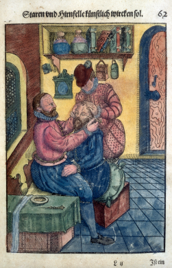
George Bartisch (1535-1607), a German physician who wrote extensively on eye disease in the 16th century, was perhaps the most well-known oculist of his time and is considered by many to be the father of modern ophthalmology (Fig. 8). He spent a considerable portion of his life plying his trade around different countries, eventually settling down in Dresden where he became court oculist to Duke Augustus I of Saxony in 1588 [27]. He is credited for publishing Ophthalmodouleia Das ist Augendienst, the first Renaissance manuscript on ophthalmic disorders and eye surgery in 1583 [28]. This comprehensive work featured 92 full-page woodcuts depicting eye disease, surgical methodology and instrumentation. It remains a pivotal work of contemporary ophthalmology and is considered by some the precursor to the modern textbook. In addition to his highly influential work, Bartisch was also the first to perform a complete eye removal in a living patient in which he removed a cancerous eye with a sharp enucleation spoon [27].
In 1722, Saint Yves published a treatise of eye pathologies that became an established pillar of the French school of ophthalmology. John Taylor (1703-1772), an English oculist and self-proclaimed Chevalier, was a controversial figure and was known as a quack having practised his art on an unsuspecting public and surgically blinding many prominent figures of his time including Handel and Bach [29]. Nonetheless, he is credited with providing one of the most complete pre-ophthalmoscopic descriptions of glaucoma. Along with John Woolhouse (1666-1734), a fellow English oculist, they recognized that glaucoma destroys the visual system [30].
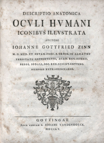
The 18th century also met the likes of Johann Gottfried Zinn (1727-1759), a German anatomist who is often referred to as the father of ocular anatomy [31]. Despite dying at the young age of 31, he left behind an incredibly vast amount of original work. In his book Descriptio Anatomica Oculi Humani (Fig. 9), Zinn provided the first detailed, comprehensive, layer-by-layer anatomy of various ocular structures [32]. His legacy is reflected by the many anatomical structures named after him, with notable examples including the zonule of Zinn, annulus of Zinn and circle of Zinn. Prominent successors who continued his work included Hippolyte Cloquet (1787-1840) and Friedrich Schlemm (1795-1858).
Modern Era (AD 1800 to Present)
Golden Age
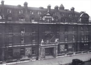
The early 19th century saw the discipline of ophthalmology becoming a well-defined area of practice due in part to the work of Austrian ophthalmologist Georg Joseph Beer (1763-1821). In 1812, he founded the first university department of ophthalmology in the general hospital of Vienna by special decree of the emperor and established the first medical school and clinic dedicated to the treatment of eye disease [33]. Ophthalmology was made a prerequisite course for medical students and Beer was appointed as the first director and professor of ophthalmology. He was also known for introducing a “flap" method (Beer’s operation) for cataract surgery and creating a special triangle-tipped instrument with an accompanying needle (Beer’s knife). Around the same time, the first dedicated ophthalmic hospital was opened in London, England in 1805 [34]. Currently known as Moorfields Eye Hospital (Fig. 10), it was initially founded as the London Dispensary for Curing Diseases of the Eye and Ear by English oculist John Cunningham Saunders (1773-1810) to care for patients suffering the blinding complications of trachoma. It is perhaps the best known ophthalmic hospital in the English-speaking world and is considered the home of British ophthalmology by many.
The mid 19th century saw the invention of the direct ophthalmoscope by Hermann von Helmholtz (1821-1894), a German physicist and physician, in 1851 which offered unprecedented diagnostic capabilities and revolutionized the practice of ophthalmology [35]. For the first time, ophthalmologists were able to unravel the mysteries of the inner eye and reveal the links between eye manifestations and systemic diseases. Albrecht von Graefe (1828-1870), a fellow German ophthalmologist (Fig. 11), further reformed modern ophthalmology and made numerous first discoveries such as excavation of the optic disc in glaucoma (1855), iridectomy in glaucoma (1857), central artery occlusion (1859) and the eponymous von Graefe’s sign seen in Graves disease [36]. He also founded the journal Archiv für Ophthalmologie (now known as Graefe’s Archive for Clinical and Experimental Ophthalmology) in 1854 and the Deutsche Ophthalmologische Gesellschaft (German Ophthalmological Society) in 1857.
Alongside von Graefe’s work, similar efforts to professionalize the field was led by Sir William Bowman (1816-1892) from Moorfields. He founded the Ophthalmological Society of the UK (now known as the Royal College of Ophthalmologists) in 1880 and provided a central organization and platform for ophthalmologists to learn through meetings and presentations [37]. An important international collaboration was also forged during the Great Exhibition of 1851 in which Bowman developed a close partnership with Albrecht von Graefe from Berlin and Frans Donders from Utrecht [38]. Bowman contributed hugely to the scientific development of ophthalmology, while von Graefe and Donders revolutionized the understanding of surgical treatment and optical science respectively.
The 20th Century
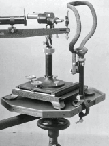
Allvar Gullstrand (1862-1930), a Swedish ophthalmologist, invented the slit lamp in 1911 with Carl Zeiss (1816-1888) of Germany using the Nernst electric bulb [39]. This groundbreaking invention (Fig. 12), which allowed a magnified, illuminated view of the interior of the eye with a slit of light, has arguably acquired the greatest importance to the practical ophthalmologist. Gullstrand's key inventions also included the reflex-free ophthalmoscope and the schematic eye. He is the only ophthalmologist to receive the Nobel Prize for medicine through his work on physical and physiological optics [40].
Advancements in various branches of ophthalmology accelerated during the 20th century. Jules Gonin (1870-1935), a Swiss ophthalmologist, is credited with changing the landscape for retinal detachment surgery by recognizing retinal breaks as the cause (and not the consequence as it was largely believed at the time) [41]. In 1922, he pioneered the procedure of ignipuncture (a technique which involved the cauterization of the retina with a very hot pointed instrument) which became the first successful surgical treatment technique for retinal detachments. The invention of light coagulation by Gerhard Meyer-Schwickerath (1920-1992) in 1946 subsequently led to the development of laser photocoagulation for the treatment of retinal defects and represented the first application of laser in medicine [42].
The 20th century also ushered in a golden age for cataract surgery due to the contributions of pioneers, most notably Sir Harold Ridley (1906-2001) and Charles Kelman (1930-2004) [43]. Although Ridley performed the first intraocular lens implant in 1949 (Fig. 13), it was not some 30 years later that it only received the recognition and respect it deserved. Nearly two decades later, Kelman (Fig. 14) introduced phacoemulsification in 1967, the seemingly perfect complement to the intraocular lens, after being inspired by his dentist’s ultrasonic probe [44]. The combination has since revolutionized the nature of cataract surgery and transformed it into an outpatient procedure due to faster postoperative recovery and the reduced need for extended hospital stays.
For more information on the history of cataract surgery, visit this EyeWiki article.
List of Important Inventions
The following is a chronological list of landmark inventions in ophthalmology and their inventors, where known:
| Invention | Year | Inventor | Country |
|---|---|---|---|
| Keratoprosthesis | 1789 | Guillaume Pellier de Quengsy | 🇫🇷France |
| Stereoscope (precursor to synoptophore) | 1832 | Charles Wheatstone | 🇬🇧UK |
| Keratoscope | 1847 | Henry Goode | 🇬🇧UK |
| Direct ophthalmoscope | 1851 | Hermann von Helmholtz | 🇩🇪Germany |
| Surgical iridectomy | 1856 | Albrecht von Graefe | 🇩🇪Germany |
| Snellen chart | 1862 | Herman Snellen | 🇳🇱Netherlands |
| Pilocarpine | 1875 | E. Hardy
Alfred Gerrard |
🇫🇷France
🇬🇧UK |
| Local anesthesia for eye surgery (cocaine) | 1884 | Carl Koller | 🇦🇹Austria |
| Hirschberg test | 1886 | Julius Hirschberg | 🇩🇪Germany |
| Corneal transplant | 1905 | Eduard Konrad Zirm | 🇦🇹Austria |
| Slit lamp | 1911 | Allvar Gullstrand | 🇸🇪Sweden |
| Ishihara color test | 1917 | Shinobu Ishihara | 🇯🇵Japan |
| Gonioscopy | 1918 | Alexios Trantas | 🇬🇷Greece |
| Streak retinoscopy | 1927 | Jack Copeland | 🇺🇸USA |
| Binocular indirect ophthalmoscope | 1945 | Charles Schepens | 🇧🇪Belgium |
| Amsler grid | 1945 | Marc Amsler | 🇨🇭Switzerland |
| Goldmann perimetry | 1945 | Hans Goldmann | 🇦🇹Austria |
| Light photocoagulation | 1946 | Gerhard Meyer-Schwickerath | 🇩🇪Germany |
| Intraocular lens | 1949 | Harold Ridley | 🇬🇧UK |
| Goldmann applanation tonometry | 1954 | Hans Goldmann | 🇦🇹Austria |
| B-scan ultrasonography | 1958 | Gilbert Baum
Ivan Greenword |
🇺🇸USA |
| Laser photocoagulation | 1960 | Theodore Maiman | 🇺🇸USA |
| Fluorescein angiography | 1961 | Harold Novotny
David Alvis |
🇺🇸USA |
| Phacoemulsification | 1967 | Charles Kelman | 🇺🇸USA |
| Trabeculectomy | 1968 | John Cairns
Peter Watson |
🇬🇧UK |
| Pars plana vitrectomy | 1970 | Robert Machemer | 🇩🇪Germany |
| Indocyanine green angiography | 1972 | Robert Flower
Bernard Hochheimer |
🇺🇸USA |
| LogMAR chart | 1976 | Ian Bailey
Jan Lovie-Kitchen |
🇦🇺Australia |
| Radial keratotomy | 1979 | Syvatoslav Fyodorov | 🇷🇺Russia |
| Humphrey field analyzer | 1984 | Mike Patella
Anders Heijl |
🇺🇸USA
🇸🇪Sweden |
| LASIK | 1989 | Gholam A Peyman | 🇺🇸USA |
| Optical coherence tomography | 1991 | James Fujimoto
Adolf Fercher David Huang Christoph Hitzenberger Eric Swanson |
🇺🇸USA
🇦🇹Austria 🇺🇸USA 🇦🇹Austria 🇺🇸USA |
Additional Resources
[Video Credit - International Council of Ophthalmology]
References
- ↑ Harper R (1904). The Code of Hammurabi, King of Babylon, about 2250 B.C. Chicago: University of Chicago Press.
- ↑ Bryan CP (1931). The Papyrus Ebers: Translated from the German Version. New York: D Appleton and Company.
- ↑ Andersen SR (1997). The eye and its diseases in Ancient Egypt. Acta Ophthalmologica, 75: 338-344.
- ↑ ReFaey K, Quinones GC, Clifton W, Tripathi S, Quiñones-Hinojosa A (2019). The Eye of Horus: The Connection Between Art, Medicine, and Mythology in Ancient Egypt. Cureus. 11(5):e4731.
- ↑ Retief F, Stulting A, & Cilliers L (2008). The eye in antiquity. SAMJ: South African Medical Journal, 98(9), 697-700.
- ↑ Craik EM (2015). The ‘Hippocratic’ Corpus: Content and Context. London; New York: Routledge, 2015. xxxvi, 307.
- ↑ Craik EM (1998). Hippocrates: Places in Man. Oxford: Clarendon Press, 25-39, 105.
- ↑ Coxe JR (1846). The writings of Hippocrates and Galen. Epitomised from the original Latin translations. Philadelphia: Lindsay and Blakiston.
- ↑ Magnus H (1999) Ophthalmology of the ancients. Translated by Waugh RL. Vol 2. Wayenborgh, Oostende pp 461–469.
- ↑ Elizondo-Omaña RE, Guzmán-López S, García-Rodríguez Mde L (2005). Dissection as a teaching tool: past, present, and future. Anat Rec B New Anat. 285:11–15.
- ↑ Von Staden H (1989). Herophilus. Cambridge: Cambridge University Press: 200-255, 570-574.
- ↑ Raju VK (2003). Susruta of ancient India. Indian J Ophthalmol; 51:119-22.
- ↑ Bhishagratna, Kaviraj Kunjalal. An English Translation of the Sushruta Samhita: Based on Original Sanskrit Text. Varanasi, India; Chowkhamba Sanskrit Series Office: 1-105.
- ↑ Grzybowski A and Ascaso FJ (2014), Sushruta in 600 B.C. introduced extraocular expulsion of lens material. Acta Ophthalmologica, 92: 194-197.
- ↑ (1994), Chapter X: The Roman Empire. Acta Ophthalmologica, 72: 76-80.
- ↑ Duke-Elder S (1961): The history of the anatomy of the eye. In: Duke- Elder S (ed.). System of ophthalmology. Vol 11, p 7-17, 20, 28, 29. The history. Vol VII, p468-469, 1962. H. Kimpton, London.
- ↑ Lazzeri D, Agostini T, Figus M, Nardi M, Spinelli G, Pantaloni M, Lazzeri S (2012). The contribution of Aulus Cornelius Celsus (25 B.C.-50 A.D.) to eyelid surgery. Orbit. 31(3):162-7.
- ↑ Leffler CT, Hadi TM, Udupa A, Schwartz SG, & Schwartz D (2016). A medieval fallacy: the crystalline lens in the center of the eye. Clinical Ophthalmology, 10, 649–662.
- ↑ Sobotka H (1957). Ophthalmology During the Middle Ages. AMA Arch Ophthalmol. 57(3):366–375.
- ↑ Sorsby A (1933). A Short History of Ophthalmology. London: Staples Press.
- ↑ Daneshfard B, Dalfardi B, Nezhad GSM (2016). Ibn al-Haytham (965–1039 AD), the original portrayal of the modern theory of vision. Journal of Medical Biography. 24(2): 227-231.
- ↑ Hirschberg J (1905). Arabian ophthalmology. J Am Med Assoc. XLV: 1127–31.
- ↑ Duke-Elder S & Wybar KC (1961). The history of the anatomy of the eye. System of Ophthalmology. Vol 2. The Anatomy of the Visual System. London: Henry Kimpton 1–72.
- ↑ De Laey JJ. The eye of Vesalius. Acta Ophthalmol. 2011 May;89(3):293-300.
- ↑ Hirschberg J (1985). The Renaissance of Ophthalmology in the 18th Century (Part 3). The First Half of the 19th Century (Part 1). The History of Ophthalmology, Vol. 5. Transl. by Blodi FC. Bonn: JP Wayenborgh 28–34.
- ↑ Loudon SE (2005). Charles de Saint-Yves (1677–1736), Strabismus, 13:3, 143-144.
- ↑ Jump up to: 27.0 27.1 Tower P (1956). Notes on the life and work of George Bartisch. AMA Arch Ophthalmol. 56(1): 57-70.
- ↑ Thompson HS (1997). Augendienst (Ophthalmodouleia or The Service of the Eyes). Arch Ophthalmol. 115(2):296.
- ↑ Wade NJ (2008). Chevalier John Taylor, ophthalmiater. Perception. 37(7): 969-72.
- ↑ Schwartz SG, Leffler CT, Grzybowski A, Koch HR, Bermudez D (2015). The Taylor Dynasty: three generations of 18th-19th century oculists. Historia Ophthalmologica Internationalis. 1: 67–81.
- ↑ Streng B, Ruprecht KW, Wittern R (1991). Johann Gottfried Zinn--ein fränkischer Anatom und Botaniker [Johann Gottfried Zinn--a Franconian anatomist and botanist]. Klin Monbl Augenheilkd. 199(1): 57-61. [German]
- ↑ Schmidt D (2019). Die bedeutende Publikation von Johann Gottfried Zinn (1727 – 1759), Descriptio anatomica oculi humani“ (1755) [The Important Publication of Johann Gottfried Zinn (1727 - 1759) "Descriptio anatomica oculi humani" (1755)]. Klin Monbl Augenheilkd.
- ↑ Albert DM, Blodi FC (1988). Georg Joseph Beer: a review of his life and contributions. Doc Ophthalmol. 68(1-2): 79-103.
- ↑ Trevor-Roper PD (1976). The History and Traditions of the Moorfields Eye Hospital. Proc R Soc Med. 69(4):316.
- ↑ Keeler CR (2002). The Ophthalmoscope in the Lifetime of Hermann von Helmholtz. Arch Ophthalmol. 120(2):194–201.
- ↑ Rohrbach JM (2020). Albrecht von Graefe in the present, the past, and the future. Graefes Arch Clin Exp Ophthalmol. 258; 1141–1147.
- ↑ Trevor-Roper P (1992). Sir William Bowman - 1816-1892. Br J Ophthalmol. 76(3):129.
- ↑ Doria C (2021). Albrecht von Graefe and the foundation of scientific ophthalmology. Indian J Ophthalmol. 69(2):211-212.
- ↑ Ehinger B, Grzybowski A (2011). Allvar Gullstrand (1862-1930) - the gentleman with the lamp. Acta Ophthalmol. 89(8):701-8.
- ↑ Ravin JG (1999). Gullstrand, Einstein, and the Nobel Prize. Arch Ophthalmol. 117(5):670–672.
- ↑ Rumpf J (1976). Jules Gonin. Inventor of the surgical treatment for retinal detachment. Surv Ophthalmol. 21(3):276-84.
- ↑ Meyer-Schwickerath GR (1989). The history of photocoagulation. Aust N Z J Ophthalmol. 17(4):427-34.
- ↑ Leffler CT, Klebanov A, Samara WA, Grzybowski A (2020). The history of cataract surgery: from couching to phacoemulsification. Ann Transl Med. 8(22):1551.
- ↑ Kelman CD (1994). The history and development of phacoemulsification. Int Ophthalmol Clin. 34(2):1-12.


