Glycogen Storage Disease Type V - McArdle Disease
All content on Eyewiki is protected by copyright law and the Terms of Service. This content may not be reproduced, copied, or put into any artificial intelligence program, including large language and generative AI models, without permission from the Academy.
This page addresses the possible ophthalmologic manifestations of glycogen storage disease type V, a rare metabolic disorder.
Glycogen storage disease type V
Disease Entity
Glycogen storage disease type V (GSD5) , also known as McArdle's disease [1], is a metabolic disorder caused by a deficiency of myophosphorylase, the muscle enzyme of glycogen phosphorylase. Its incidence is reported as one in 100,000 [2].
Etiology
Two autosomal recessive forms of this disease occur, childhood-onset and adult-onset. The gene for myophosphorylase, PYGM (the muscle-type of the glycogen phosphorylase gene), is located on chromosome 11q13. Myophosphorylase is involved in the breakdown of glycogen to glucose. The enzyme removes 1,4 glycosyl residues from outer branches of glycogen and adds inorganic phosphate to form glucose-1-phosphate. Cells form glucose-1-phosphate instead of glucose during glycogen breakdown because the polar, phosphorylated glucose cannot leave the cell membrane and so is marked for intracellular catabolism.
Clinical manifestations
Clinically, it manifests as exercise intolerance and acute episodes of contractures, associated with rhabdomyolysis and myoglobinuria.
Diagnosis
- A muscle biopsy will note the absence of myophosphorylase in muscle fibers. In some cases, acid-Schiff stained glycogen can be seen with microscopy.
- Genetic sequencing of the PYGM gene

Ophthalmologic manifestations
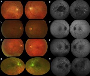
Several case-reports and case-series have associated GSD5 with a retinal pattern dystrophy [3][4][5][6]. Glucose is the major energy substrate for retinal metabolism and its source to the outer retina is the retinal pigmented epithelium (RPE) which manages the conversion of glycogen to glucose.[7] It has been demonstrated that, in human RPE, the glycogen phosphorylase enzyme present is the muscle isoform (myophosphorylase). [8] This reinforces the pathogenesis of retinopathy in McArdle disease.
References
- ↑ McArdle B. Myopathy due to a defect in muscle glycogen breakdown. Clin Sci. 1951. doi:10.5694/j.1326-5377.1951.tb74724.x
- ↑ http://mcardlesdisease.org/
- ↑ 3.0 3.1 Mahroo OA, Khan KN, Wright G, Ockrim Z, Scalco RS, Robson AG, Tufail A, Michaelides M, Quinlivan R, Webster AR. Retinopathy Associated with Biallelic Mutations in PYGM (McArdle Disease). Ophthalmology. 2019 Feb;126(2):320-322. doi: 10.1016/j.ophtha.2018.09.013. Epub 2018 Oct 11. PMID: 30316539; PMCID: PMC6347563.
- ↑ Marques JH, Beirão JM (2020) McArdle Disease associated Maculopathy and the Role of Glycogen in the Retina. J Ophthalmic Clin Res 7: 067.
- ↑ Casalino G, Chan W, McAvoy C, Coppola M, Bandello F, Bird AC, Chakravarthy U. Multimodal imaging of posterior ocular involvement in McArdle's disease. Clin Exp Optom. 2018 May;101(3):412-415. doi: 10.1111/cxo.12635. Epub 2017 Nov 26. PMID: 29178179.
- ↑ Alsberge JB, Chen JJ, Zaidi AA, Fu AD. RETINAL DYSTROPHY IN A PATIENT WITH MCARDLE DISEASE. Retin Cases Brief Rep. 2018 Aug 1. doi: 10.1097/ICB.0000000000000790. PMID: 30074569.
- ↑ Senanayake Pd, Calabro A, Hu JG, Bonilha VL, Darr A, Bok D, Hollyfield JG. Glucose utilization by the retinal pigment epithelium: evidence for rapid uptake and storage in glycogen, followed by glycogen utilization. Exp Eye Res. 2006 Aug;83(2):235-46. doi: 10.1016/j.exer.2005.10.034. Epub 2006 May 11. PMID: 16690055.
- ↑ Hernández C, Garcia-Ramírez M, García-Rocha M, Saez-López C, Valverde ÁM, Guinovart JJ, Simó R. Glycogen storage in the human retinal pigment epithelium: a comparative study of diabetic and non-diabetic donors. Acta Diabetol. 2014 Aug;51(4):543-52. doi: 10.1007/s00592-013-0549-8. Epub 2014 Jan 24. PMID: 24458975.

