File:Xu - Focal Choroidal Excavation.jpeg
Xu_-_Focal_Choroidal_Excavation.jpeg (590 × 361 pixels, file size: 89 KB, MIME type: image/jpeg)
Summary
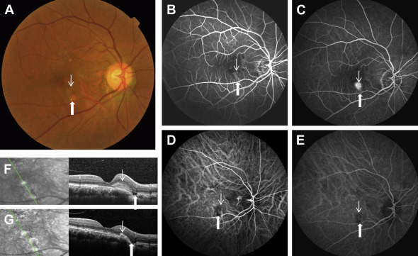
A. Color photograph of the right eye demonstrates a combined focal choroidal excavation (FCE) with choroidal neovascularization (CNV)
B and C. Fundus fluorescein angiography demonstrates the corresponding FCE and CNV
D and E. Indocyanine green angiography demonstrates the corresponding FCE and CNV
F and G. Spectral-domain optical coherence tomography demonstrates a nonconforming FCE and CNV.
G demonstrates resolution of the CNV after intravitreal injection of ranibizumab along with the nonconforming FCE turning into the conforming type.
Thin arrow corresponds to CNV, Thick arrow corresponds to FCE
(Courtesy of Haifeng Xu, MD, PhD; Source: Xu, H. et al. Focal choroidal excavation complicated by choroidal neovascularization. Ophthalmology 121, 246–250 (2014).)
File history
Click on a date/time to view the file as it appeared at that time.
| Date/Time | Thumbnail | Dimensions | User | Comment | |
|---|---|---|---|---|---|
| current | 15:15, June 19, 2022 | | 590 × 361 (89 KB) | Jane.Z.Spadaro (talk | contribs) | A. Color photograph of the right eye demonstrates a combined focal choroidal excavation (FCE) with choroidal neovascularization (CNV) B and C. Fundus fluorescein angiography demonstrates the corresponding FCE and CNV D and E. Indocyanine green angiography demonstrates the corresponding FCE and CNV F and G. Spectral-domain optical coherence tomography demonstrates a nonconforming FCE and CNV. G demonstrates resolution of the CNV after intravitreal injection of ranibizumab along with the nonco... |
You cannot overwrite this file.
File usage
The following page uses this file: