File:Sympathetics cav sinus.png
From EyeWiki

Size of this preview: 800 × 544 pixels. Other resolution: 974 × 662 pixels.
Original file (974 × 662 pixels, file size: 475 KB, MIME type: image/png)
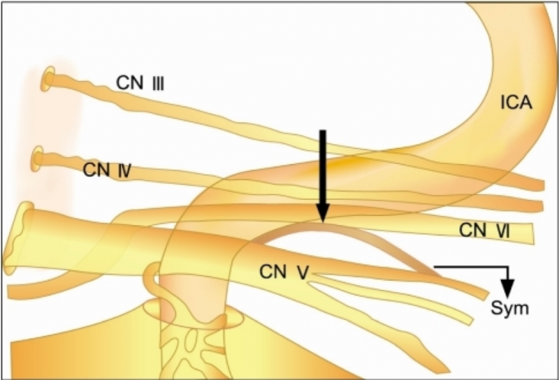
Figure 1: A depiction of the posterior portion of the cavernous sinus, demonstrating oculosympathetic fibers leaving the sympathetic plexus around the ICA and moving onto CN VI before exiting on the ophthalmic branch of CN V (V1). Copyright: Open-access image, unaltered from original. License: https://creativecommons.org/licenses/by-nc/3.0/ Original: https://openi.nlm.nih.gov/detailedresult.php?img=PMC3223717_kjo-25-459-g005&req=4
File history
Click on a date/time to view the file as it appeared at that time.
| Date/Time | Thumbnail | Dimensions | User | Comment | |
|---|---|---|---|---|---|
| current | 15:48, March 14, 2019 | | 974 × 662 (475 KB) | Bayan.Al Othman (talk | contribs) | Figure 1: A depiction of the posterior portion of the cavernous sinus, demonstrating oculosympathetic fibers leaving the sympathetic plexus around the ICA and moving onto CN VI before exiting on the ophthalmic branch of CN V (V1). Copyright: Open-acc... |
You cannot overwrite this file.
File usage
The following page uses this file: