File:Stage 3 VL.jpg
From EyeWiki
Size of this preview: 800 × 151 pixels. Other resolution: 3,245 × 611 pixels.
Original file (3,245 × 611 pixels, file size: 228 KB, MIME type: image/jpeg)
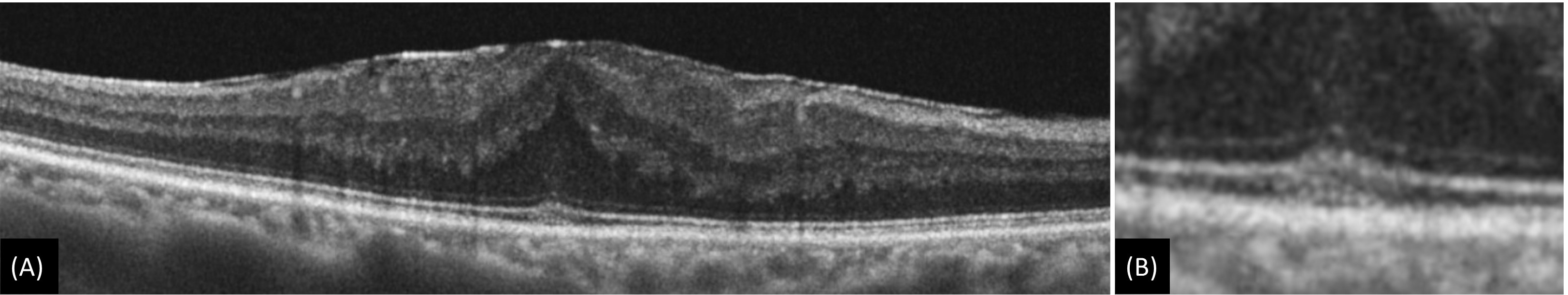
Figure 5: (A) Epiretinal membrane with acquired vitelliform lesion which is characterized by presence of thick dome-shaped subfoveal hyperreflective material between ellipsoid zone and retinal pigment epithelium. (B) Magnified image
File history
Click on a date/time to view the file as it appeared at that time.
| Date/Time | Thumbnail | Dimensions | User | Comment | |
|---|---|---|---|---|---|
| current | 11:01, May 31, 2023 | 3,245 × 611 (228 KB) | Kushal.Delhiwala (talk | contribs) |
You cannot overwrite this file.
File usage
The following page uses this file: