File:Stage 2 foveolar detachment.jpg
From EyeWiki
Size of this preview: 798 × 166 pixels. Other resolution: 3,240 × 674 pixels.
Original file (3,240 × 674 pixels, file size: 202 KB, MIME type: image/jpeg)
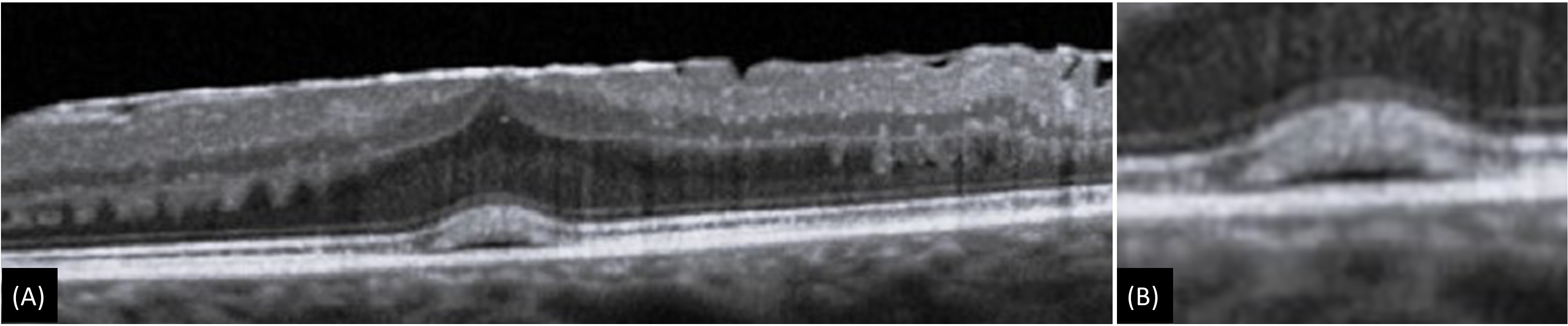
Figure 4: (A) Epiretinal membrane with foveolar detachment which is characterized by central subfoveal hyporeflective pocket corresponding to subretinal fluid accumulation under interdigitation zone. (B) Magnified image
File history
Click on a date/time to view the file as it appeared at that time.
| Date/Time | Thumbnail | Dimensions | User | Comment | |
|---|---|---|---|---|---|
| current | 11:00, May 31, 2023 | 3,240 × 674 (202 KB) | Kushal.Delhiwala (talk | contribs) |
You cannot overwrite this file.
File usage
The following page uses this file: