File:Screenshot 2023-05-30 at 10.17.24 AM.png
Screenshot_2023-05-30_at_10.17.24_AM.png (640 × 484 pixels, file size: 624 KB, MIME type: image/png)
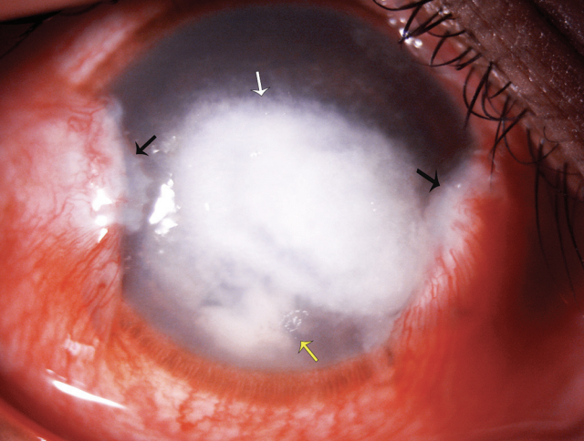
- Slit‑lamp image of double pterygium with pythium keratitis. The
The image depicts nasal and temporal pterygium (black arrowheads), central full‑thickness corneal infiltrate with tentacular projections (white arrowhead), and the tendency for early limbal spread (yellow arrowhead). These features are the hallmark of pythium keratitis, which help in distinguishing it clinically from fungal keratitis.
Reproduced by permission from Editor in Chief- TJOSR
Source-Gurnani, Bharat; Narayana, Shivanand; Christy, Josephine; Kaur, Kirandeep. Double Pterygium with Pythium Keratitis. TNOA Journal of Ophthalmic Science and Research 59(2):p 225, Apr–Jun 2021. | DOI: 10.4103/tjosr.tjosr_183_20
File history
Click on a date/time to view the file as it appeared at that time.
| Date/Time | Thumbnail | Dimensions | User | Comment | |
|---|---|---|---|---|---|
| current | 21:51, May 29, 2023 | | 640 × 484 (624 KB) | Bharat.Gurnani (talk | contribs) |
You cannot overwrite this file.
File usage
The following page uses this file: