File:Screening Figure 1.png
From EyeWiki

Size of this preview: 800 × 518 pixels. Other resolution: 1,280 × 828 pixels.
Original file (1,280 × 828 pixels, file size: 2.2 MB, MIME type: image/png)
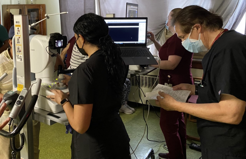
Figure 1. SSSP Volunteers using non-mydriatic retinal cameras to capture images of a patient’s anterior segment and posterior pole (retina), while certified reader Dr. Bernard Szirth interprets patient data. Note subjects as well as imagers all stand at the instruments to streamline the process. When subjects sit at an instrument, the process can take 40% longer. Especially in times of COVID-19, it is important to minimize time at each data collecting station to limit exposure.
File history
Click on a date/time to view the file as it appeared at that time.
| Date/Time | Thumbnail | Dimensions | User | Comment | |
|---|---|---|---|---|---|
| current | 13:06, September 27, 2021 | | 1,280 × 828 (2.2 MB) | Albertkhouri (talk | contribs) |
You cannot overwrite this file.
File usage
The following page uses this file: