File:Screen Shot 2023-04-27 at 7.31.09 PM.png
From EyeWiki
Size of this preview: 800 × 450 pixels. Other resolution: 1,646 × 926 pixels.
Original file (1,646 × 926 pixels, file size: 2.03 MB, MIME type: image/png)
Summary
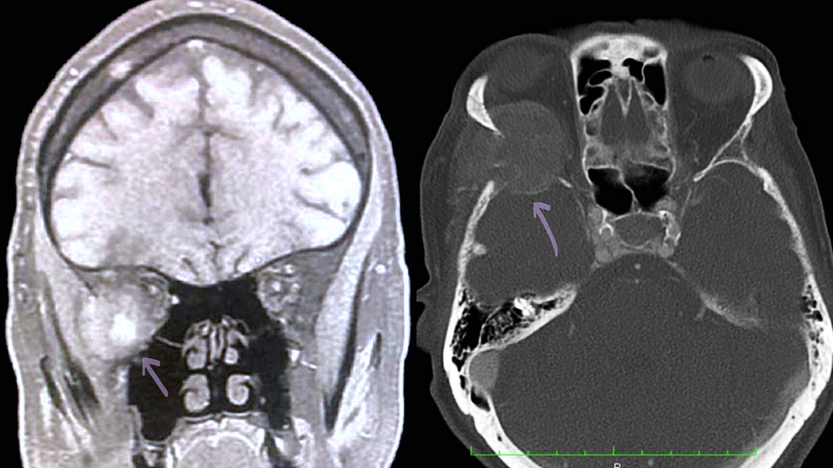
Orbital Pheochromocytoma MRI versus CT (Left) Coronal MRI without contrast showing a heterogeneously enhancing mass in the right orbit. (Right) Axial CT orbit with contrast revealing a orbital lesion involving the bony and orbital tissue.
Rider AJ, Walsh A, Sollenberger EL, Dryden SC, DeAngelis KD, Weir AB 3rd, Fowler BT. Orbital Pheochromocytoma Metastasis in 2 Patients With Known Pheochromocytoma. Ophthalmic Plast Reconstr Surg. 2019 Nov/Dec;35(6):e131-e134. doi: 10.1097/IOP.0000000000001460. PMID: 31593045.
File history
Click on a date/time to view the file as it appeared at that time.
| Date/Time | Thumbnail | Dimensions | User | Comment | |
|---|---|---|---|---|---|
| current | 17:30, April 27, 2023 | | 1,646 × 926 (2.03 MB) | Fabliha.Anbar (talk | contribs) | Orbital Pheochromocytoma MRI versus CT (Left) Coronal MRI without contrast showing a heterogeneously enhancing mass in the right orbit. (Right) Axial CT orbit with contrast revealing a orbital lesion involving the bony and orbital tissue. Rider AJ, Walsh A, Sollenberger EL, Dryden SC, DeAngelis KD, Weir AB 3rd, Fowler BT. Orbital Pheochromocytoma Metastasis in 2 Patients With Known Pheochromocytoma. Ophthalmic Plast Reconstr Surg. 2019 Nov/Dec;35(6):e131-e134. doi: 10.1097/IOP.0000000000001... |
You cannot overwrite this file.
File usage
The following page uses this file: