File:Screen Shot 2023-04-27 at 7.28.31 PM.png
Original file (1,958 × 506 pixels, file size: 2.16 MB, MIME type: image/png)
Summary
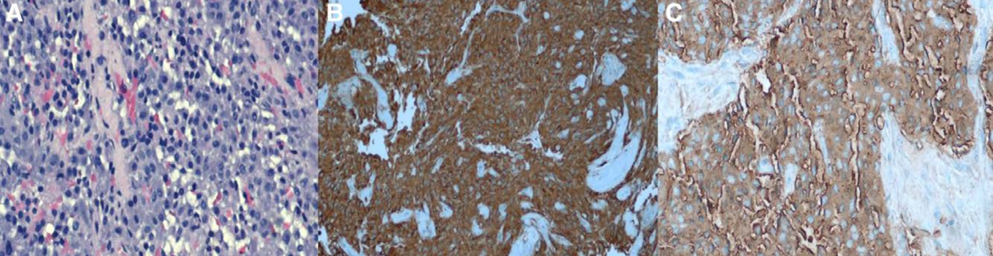
Histopathology of Metastatic Orbital Pheochromocytoma - (Left) 20x magnification with small irregular nests and clusters separated by vascular stroma with atypical nuclear features. (Center) Synaptophysin IHC strongly positive in tumor cells. (Right) Chromogranin IHC strongly positive staining in tumor cells.
Rider AJ, Walsh A, Sollenberger EL, Dryden SC, DeAngelis KD, Weir AB 3rd, Fowler BT. Orbital Pheochromocytoma Metastasis in 2 Patients With Known Pheochromocytoma. Ophthalmic Plast Reconstr Surg. 2019 Nov/Dec;35(6):e131-e134. doi: 10.1097/IOP.0000000000001460. PMID: 31593045.
File history
Click on a date/time to view the file as it appeared at that time.
| Date/Time | Thumbnail | Dimensions | User | Comment | |
|---|---|---|---|---|---|
| current | 17:32, April 27, 2023 | 1,958 × 506 (2.16 MB) | Fabliha.Anbar (talk | contribs) | Histopathology of Metastatic Orbital Pheochromocytoma - (Left) 20x magnification with small irregular nests and clusters separated by vascular stroma with atypical nuclear features. (Center) Synaptophysin IHC strongly positive in tumor cells. (Right) Chromogranin IHC strongly positive staining in tumor cells. Rider AJ, Walsh A, Sollenberger EL, Dryden SC, DeAngelis KD, Weir AB 3rd, Fowler BT. Orbital Pheochromocytoma Metastasis in 2 Patients With Known Pheochromocytoma. Ophthalmic Plast Re... |
You cannot overwrite this file.
File usage
The following page uses this file: