File:Screen Shot 2022-01-30 at 10.05.41 AM.png
Screen_Shot_2022-01-30_at_10.05.41_AM.png (674 × 412 pixels, file size: 195 KB, MIME type: image/png)
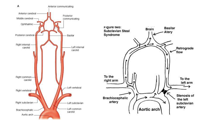
Image 1A and 1B | Blood flow in the Vertebral Artery
In the images above, Image 1A demonstrates the blood flow in the vertebral artery when there is no subclavian stenosis. As one can note, the flow is not impeded nor is there any pressure gradient favorability. On the other hand, Image 1B demonstrates the blood flow in the vertebral artery when there is subclavian stenosis. From the image provided, the blood flow is affected. This results in retrograde blood flow in the vertebral artery or internal thoracic artery, which is due to proximal stenosis/occlusion of the subclavian artery.
File history
Click on a date/time to view the file as it appeared at that time.
| Date/Time | Thumbnail | Dimensions | User | Comment | |
|---|---|---|---|---|---|
| current | 09:06, January 30, 2022 | | 674 × 412 (195 KB) | Chaow.Charoenkijkajorn (talk | contribs) |
You cannot overwrite this file.
File usage
The following page uses this file: