File:RDD Pathology.png
Original file (974 × 889 pixels, file size: 1.8 MB, MIME type: image/png)
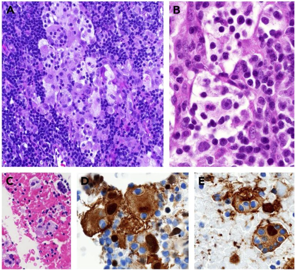
Figure 2. Nodal RDD from tissue biopsies (A-B) and fine-needle aspiration (C-E). (A) Mixed RDD/LCH case with sinus expansion. The large RDD histiocytes display conspicuous emperipolesis with pale cytoplasm, as compared with the intermixed LCH cells with dense eosinophilic cytoplasm and convoluted nuclei (OM ×400; H&E stain). (B) The RDD histiocytes show pale watery-clear cytoplasm, a central round nucleus with a conspicuous nucleolus, and emperipolesis (OM ×1000; H&E stain). Cell block preparation shows clusters of RDD histiocytes (OM ×400; H&E stain) (C), with nuclear and cytoplasmic staining for S100 (OM ×1000) (D) and fascin (OM ×1000) (E); the trafficking intact leukocytes are negative. (11)
File history
Click on a date/time to view the file as it appeared at that time.
| Date/Time | Thumbnail | Dimensions | User | Comment | |
|---|---|---|---|---|---|
| current | 16:05, September 14, 2020 | | 974 × 889 (1.8 MB) | Alexander.Engelmann (talk | contribs) | Figure 2. Nodal RDD from tissue biopsies (A-B) and fine-needle aspiration (C-E). (A) Mixed RDD/LCH case with sinus expansion. The large RDD histiocytes display conspicuous emperipolesis with pale cytoplasm, as compared with the intermixed LCH cells wit... |
You cannot overwrite this file.
File usage
The following page uses this file: