File:RDD CT Orbits.png
From EyeWiki
Size of this preview: 475 × 600 pixels. Other resolution: 750 × 947 pixels.
Original file (750 × 947 pixels, file size: 840 KB, MIME type: image/png)
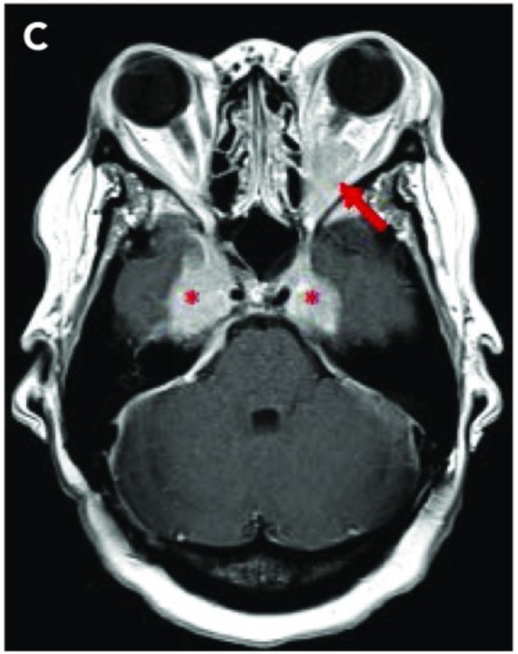
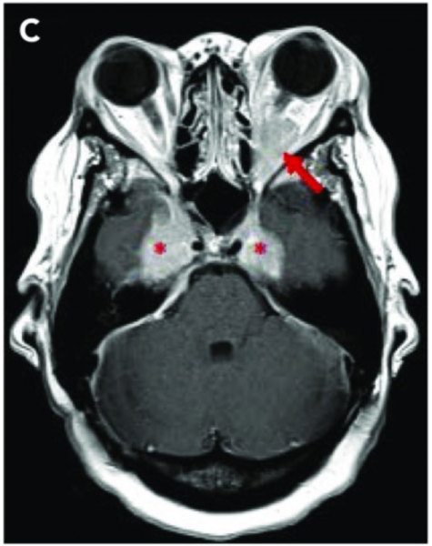
Figure 3. Gadolinium enhanced axial T1 weighted imaging demonstrates lesions marked with asterisks in bilateral cavernous sinuses and Meckel’s cave. Also an arrow points to left intraconal lesion causing proptosis (12).
File history
Click on a date/time to view the file as it appeared at that time.
| Date/Time | Thumbnail | Dimensions | User | Comment | |
|---|---|---|---|---|---|
| current | 16:41, September 14, 2020 | | 750 × 947 (840 KB) | Alexander.Engelmann (talk | contribs) | Reverted to version as of 23:35, September 14, 2020 |
| 16:41, September 14, 2020 | Error creating thumbnail: File missing | 750 × 947 (840 KB) | Alexander.Engelmann (talk | contribs) | ||
| 16:35, September 14, 2020 | Error creating thumbnail: File missing | 750 × 947 (840 KB) | Alexander.Engelmann (talk | contribs) | Figure 3. Gadolinium enhanced axial T1 weighted imaging demonstrates lesions marked with asterisks in bilateral cavernous sinuses and Meckel’s cave. Also an arrow points to left intraconal lesion causing proptosis (12). |
You cannot overwrite this file.
File usage
The following page uses this file: