File:Posterior Vortex Vein or Macular Vortex Vein.png
Original file (3,772 × 2,328 pixels, file size: 6.13 MB, MIME type: image/png)
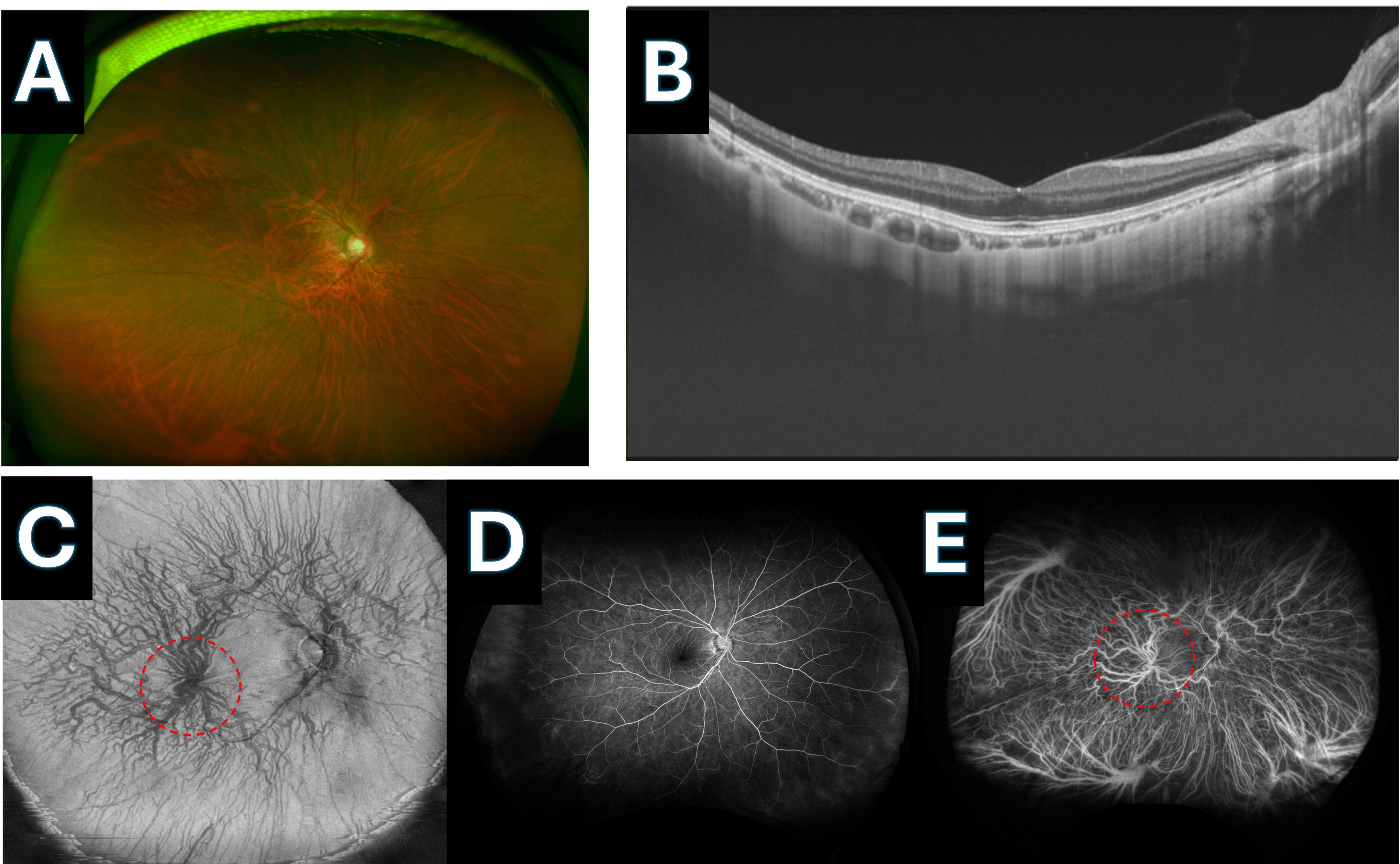
Posterior Vortex Vein or Macular Vortex Vein A: Widefield fundus image showing thinning of the retina, with clearly visible choroidal vessels through the retina, and an evident posterior vortex vein. B: Image depicting large hyperreflective areas deep to the choroid, corresponding to larger vortex vein branches and the vortex vein ampulla. C: Ultra-widefield enface swept-source optical coherence tomography (SS-OCT) image of the right eye, clearly showing the vortex vein ampulla circled by the red dotted line in the posterior pole. D: Ultra-widefield fluorescein angiography (UWFA) (2 minutes 36 seconds) revealing no retinal vascular abnormalities in the posterior pole. E: Ultra-widefield indocyanine green fluorescence angiography (UWICGA) (2 minutes). [1]
- ↑ Nitta K, Akiyama H (July 02, 2024) Different Vortex Vein Anomalies Observed in a Single Case: Macular Vortex Vein in One Eye and Varix of Vortex Vein Ampulla in the Other Eye. Cureus 16(7): e63668. doi:10.7759/cureus.63668
File history
Click on a date/time to view the file as it appeared at that time.
| Date/Time | Thumbnail | Dimensions | User | Comment | |
|---|---|---|---|---|---|
| current | 11:40, August 31, 2024 | | 3,772 × 2,328 (6.13 MB) | Joobin.Khadamy (talk | contribs) |
You cannot overwrite this file.
File usage
The following page uses this file: