File:Pearce Figure 1b.jpg
From EyeWiki

Size of this preview: 697 × 599 pixels. Other resolutions: 2,382 × 2,048 pixels | 2,615 × 2,248 pixels.
Original file (2,615 × 2,248 pixels, file size: 1.25 MB, MIME type: image/jpeg)
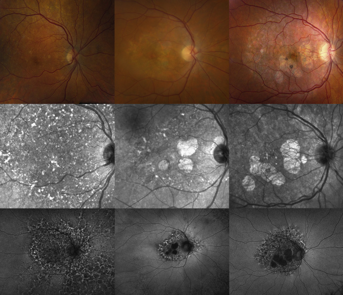
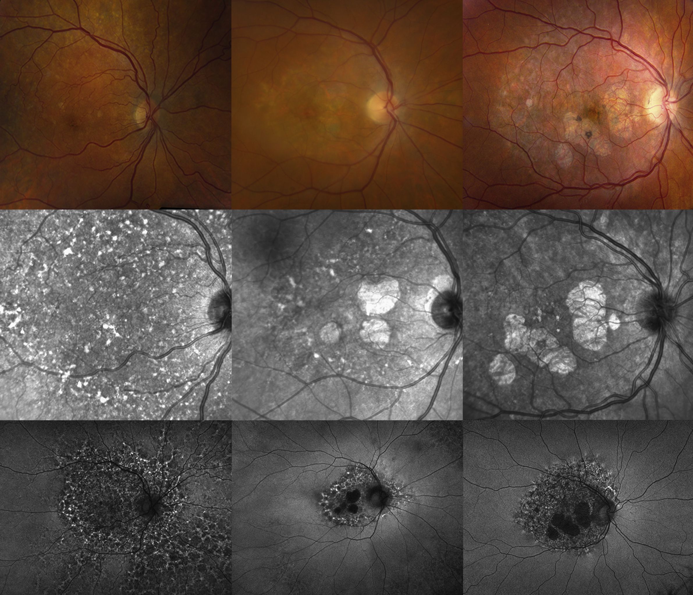
Disease spectrum in pentosan polysulfate maculopathy shown via color fundus photography (top row), near infrared reflectance imaging (middle row), and fundus autofluorescent imaging (bottom row). Part 2/2 of this complete figure.
File history
Click on a date/time to view the file as it appeared at that time.
| Date/Time | Thumbnail | Dimensions | User | Comment | |
|---|---|---|---|---|---|
| current | 13:23, January 22, 2021 | | 2,615 × 2,248 (1.25 MB) | Nieraj.Jain (talk | contribs) | Disease spectrum in pentosan polysulfate maculopathy shown via color fundus photography (top row), near infrared reflectance imaging (middle row), and fundus autofluorescent imaging (bottom row). Part 2 of 2 of this figure. Reprinted with permission fr... |
| 13:23, January 22, 2021 |  | 2,615 × 2,248 (1.25 MB) | Nieraj.Jain (talk | contribs) | Disease spectrum in pentosan polysulfate maculopathy shown via color fundus photography (top row), near infrared reflectance imaging (middle row), and fundus autofluorescent imaging (bottom row). Part 2 of 2 of this figure. Reprinted with permission fr... | |
| 06:34, January 22, 2021 |  | 2,615 × 2,248 (1.25 MB) | Nieraj.Jain (talk | contribs) | Disease spectrum in pentosan polysulfate maculopathy shown via color fundus photography (top row), near infrared reflectance imaging (middle row), and fundus autofluorescent imaging (bottom row). Part 2/2 of this complete figure. |
You cannot overwrite this file.
File usage
The following page uses this file: