File:Pathophysiology of CB abnormalities.jpg
From EyeWiki
Size of this preview: 800 × 328 pixels. Other resolution: 3,507 × 1,436 pixels.
Original file (3,507 × 1,436 pixels, file size: 626 KB, MIME type: image/jpeg)
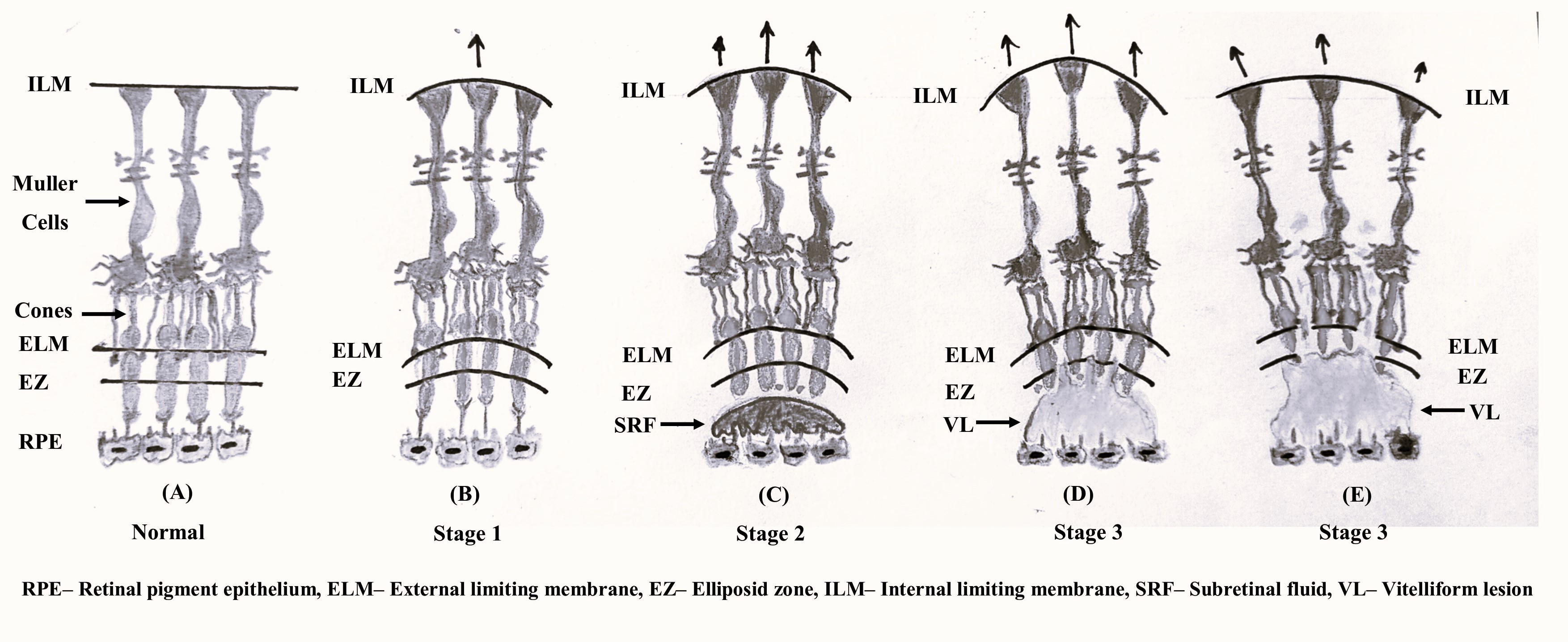
Figure 2: Schematic drawing showing (A) Arrangement of Muller cell cone in relation to cone photoreceptors at central fovea. Transmission of mechanical stress from Muller cells to cones leads to progressive development of stage 1 (B), stage 2 (C) and stage 3 (D,E) of central bouquet abnormalities. Art work in the drawing inspired from Govetto et al [1] and Salman et al [15].
File history
Click on a date/time to view the file as it appeared at that time.
| Date/Time | Thumbnail | Dimensions | User | Comment | |
|---|---|---|---|---|---|
| current | 10:57, May 31, 2023 | 3,507 × 1,436 (626 KB) | Kushal.Delhiwala (talk | contribs) |
You cannot overwrite this file.
File usage
The following page uses this file: