File:PCV.jpg
From EyeWiki
No higher resolution available.
PCV.jpg (643 × 363 pixels, file size: 138 KB, MIME type: image/jpeg)
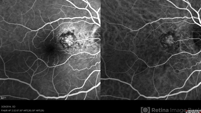
Polypoidal choroidal vasculopathy - The PCV lesion shows hyperfluorescence and hypercyanescence on intermediate-phase FA (left) and ICGA (right). This image was originally published in the Retina Image Bank® website. Gareth Lema MD, PhD. Photographer Sandra Boglione.Polypoidal Choroidal Vasculopathy - IVFA/ICGA. Retina Image Bank. 2018; 28352. © the American Society of Retina Specialists.
File history
Click on a date/time to view the file as it appeared at that time.
| Date/Time | Thumbnail | Dimensions | User | Comment | |
|---|---|---|---|---|---|
| current | 10:33, November 23, 2023 | | 643 × 363 (138 KB) | Evan.Wotipka (talk | contribs) |
You cannot overwrite this file.
File usage
The following page uses this file: