File:Orbital implant 14.jpg
Orbital_implant_14.jpg (540 × 547 pixels, file size: 103 KB, MIME type: image/jpeg)
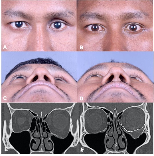
Figure 14: All the images belong to the same patient. (A and B) pre and post-operative images in the standard view with pre-operative image demonstrating left superior sulcus deformity and the post-operative image suggestive of decreased superior sulcus deformity. (C and D) Pre and post-operative image in the Bird’s view demonstrating minimal reduction in proptosis, (E and F) per and post-operative image of coronal CT section, in bone window with the pre-operative image showing floor and medial wall fracture and the post-operative image demonstrating a titanium mesh along the medial wall and floor.
File history
Click on a date/time to view the file as it appeared at that time.
| Date/Time | Thumbnail | Dimensions | User | Comment | |
|---|---|---|---|---|---|
| current | 00:30, June 1, 2021 | | 540 × 547 (103 KB) | Gangadhara.Sundar (talk | contribs) |
You cannot overwrite this file.
File usage
The following page uses this file: