File:OCT mac wagner.jpg
Original file (1,577 × 1,050 pixels, file size: 244 KB, MIME type: image/jpeg)
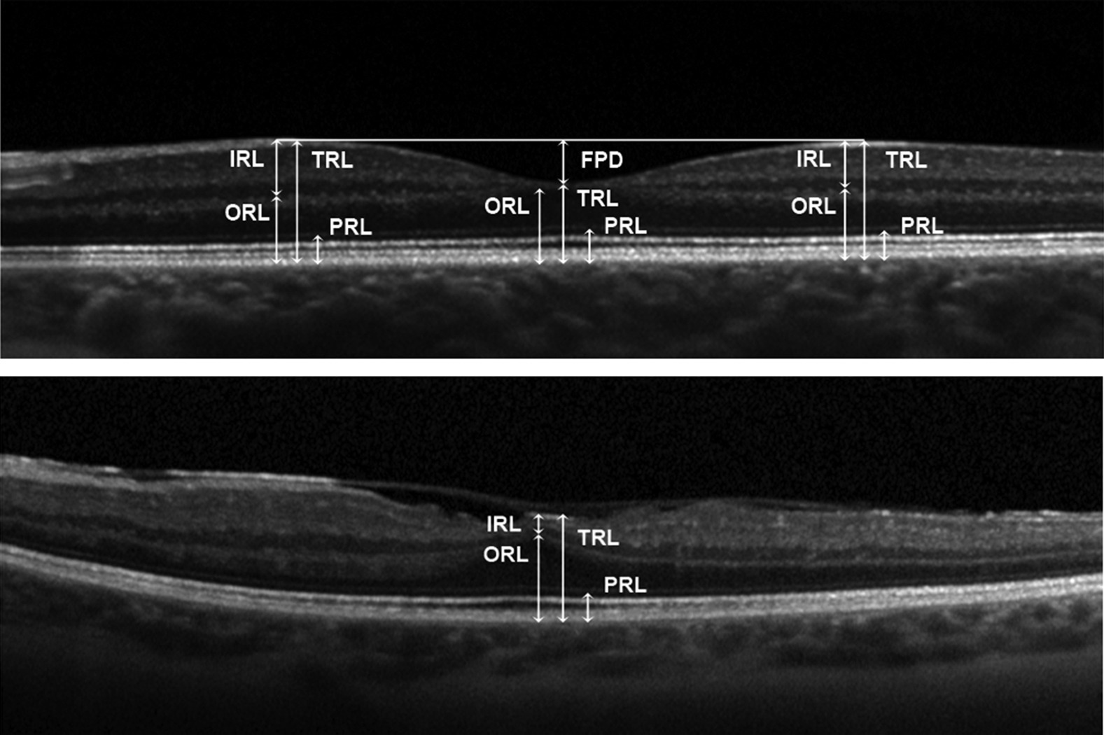
Point estimates of the thickness of several retinal layers in control subjects (Top) and in patients with Wagner syndrome (Bottom). Measures of the total retinal thickness (TRT), inner retinal layers (IRL), outer retinal layers (ORL), and photoreceptor layer (PRL) as well as the foveal pit depth (FPD) were performed in the center of the fovea and at 1000 μm of eccentricity along the horizontal line scan. Ref: Rothschild PR, Burin-des-Roziers C, Audo I, Nedelec B, Valleix S, Brézin AP. Spectral-Domain Optical Coherence Tomography in Wagner Syndrome: Characterization of Vitreoretinal Interface and Foveal Changes. Am J Ophthalmol. 2015 Nov;160(5):1065-1072.e1.
File history
Click on a date/time to view the file as it appeared at that time.
| Date/Time | Thumbnail | Dimensions | User | Comment | |
|---|---|---|---|---|---|
| current | 13:18, September 10, 2023 | | 1,577 × 1,050 (244 KB) | Janine.Collinge (talk | contribs) |
You cannot overwrite this file.
File usage
The following page uses this file: