File:Morbihan Pathology.jpg
From EyeWiki

Size of this preview: 800 × 533 pixels. Other resolutions: 2,560 × 1,706 pixels | 3,209 × 2,139 pixels.
Original file (3,209 × 2,139 pixels, file size: 1.94 MB, MIME type: image/jpeg)
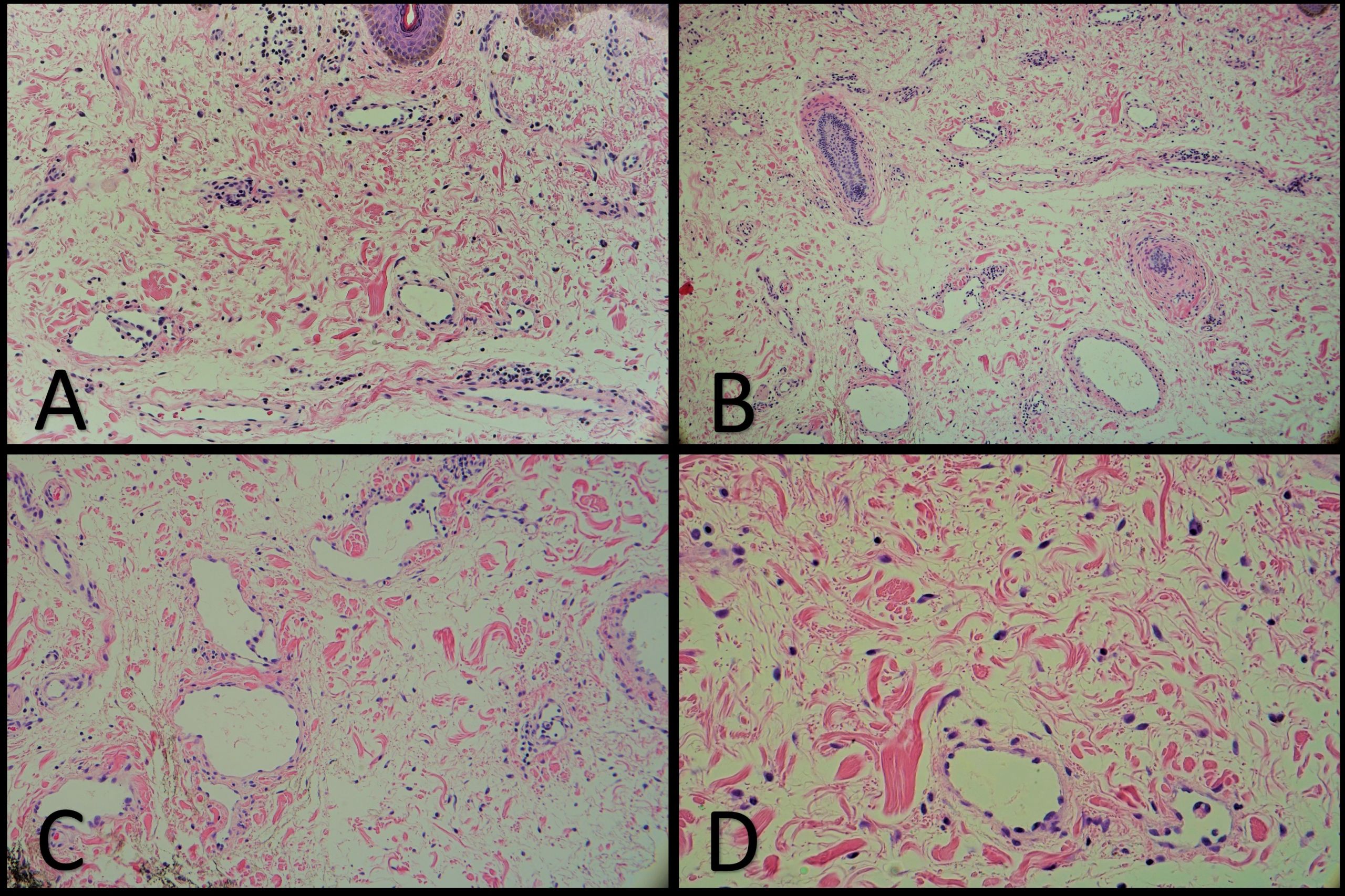
Histopathology of upper eyelid tissue in a patient with Morbihan disease using hematoxylin and eosin (H&E) stain. Findings are non-specific. (A) 10x. Dermal edema with dilated lymphatics. (B) 10x. Perifollicular fibrosis, dermal edema, and dilated vessels. (C) 20x. Dilated lymphatics and dermal edema. (D) 40x. Dermal edema and mast cells. Slides provided by David Plemel, MD.
File history
Click on a date/time to view the file as it appeared at that time.
| Date/Time | Thumbnail | Dimensions | User | Comment | |
|---|---|---|---|---|---|
| current | 21:07, September 1, 2021 | | 3,209 × 2,139 (1.94 MB) | David.Plemel (talk | contribs) |
You cannot overwrite this file.
File usage
The following page uses this file: