File:Medulloepithelioma Mass Iris.jpeg
From EyeWiki
No higher resolution available.
Medulloepithelioma_Mass_Iris.jpeg (512 × 409 pixels, file size: 31 KB, MIME type: image/jpeg)
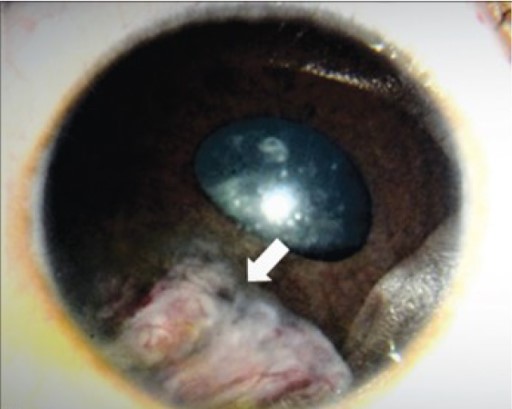
Figure - A fleshy vascularized mass is visualized in the eye, confirmed on fine needle aspiration biopsy to have ciliary body medulloepithelioma with angle involvement. (Adapted with permission from Honavar et al. 2019 Indian Journal of Ophthalmology)
File history
Click on a date/time to view the file as it appeared at that time.
| Date/Time | Thumbnail | Dimensions | User | Comment | |
|---|---|---|---|---|---|
| current | 18:37, November 15, 2019 | | 512 × 409 (31 KB) | Mohammad.Sadiq (talk | contribs) | Figure - A fleshy vascularized mass is visualized in the eye, confirmed on fine needle aspiration biopsy to have ciliary body medulloepithelioma with angle involvement. (Adapted with permission from Honavar et al. 2019 Indian Journal of Ophthalmology) |
You cannot overwrite this file.
File usage
The following page uses this file: