File:Mcardle fundus.jpg

Original file (769 × 648 pixels, file size: 156 KB, MIME type: image/jpeg)
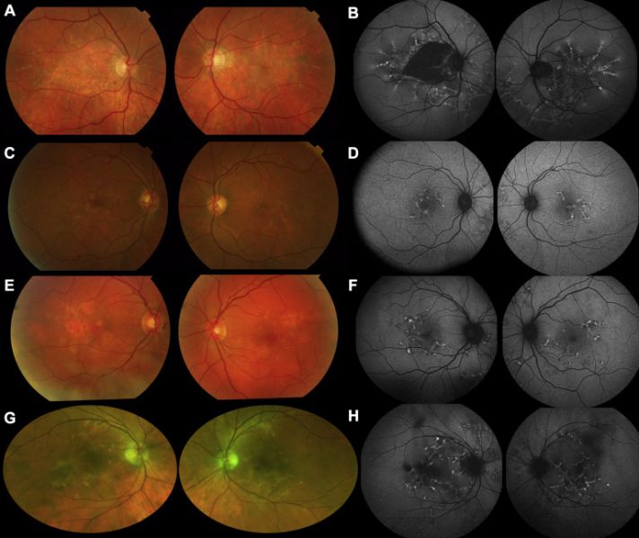
Color imaging (left panels) and fundus autofluorescence (FAF) imaging (right panels). A, Color fundus images from patient 1. B, Short-wavelength (488-nm) posterior pole (55°) FAF images from patient 1. C, D, Corresponding images from patient 2. E, F, Corresponding images from patient 3. G, H, Pseudocolor image and 488-nm FAF image from patient 4. In all cases, yellow reticular lesions are visible on color imaging, with atrophy in patient 1 and small areas of pigmentary change in patients 2, 3, and 4. Fundus autofluorescence (FAF) shows reticular hyperautofluorescence (likely to result from loss of photoreceptor outer segments, accumulation of fluorophore in the retinal pigment epithelium [RPE], or both) in all cases, with additional dark areas indicating RPE atrophy in patient 1.
From: Mahroo OA, Khan KN, Wright G, Ockrim Z, Scalco RS, Robson AG, Tufail A, Michaelides M, Quinlivan R, Webster AR. Retinopathy Associated with Biallelic Mutations in PYGM (McArdle Disease). Ophthalmology. 2019 Feb;126(2):320-322. doi: 10.1016/j.ophtha.2018.09.013.
File history
Click on a date/time to view the file as it appeared at that time.
| Date/Time | Thumbnail | Dimensions | User | Comment | |
|---|---|---|---|---|---|
| current | 15:55, January 21, 2023 | | 769 × 648 (156 KB) | Yicheng.Bao (talk | contribs) |
You cannot overwrite this file.
File usage
The following page uses this file: