File:Koizumi SlitLamp.png
Original file (736 × 979 pixels, file size: 1,013 KB, MIME type: image/png)
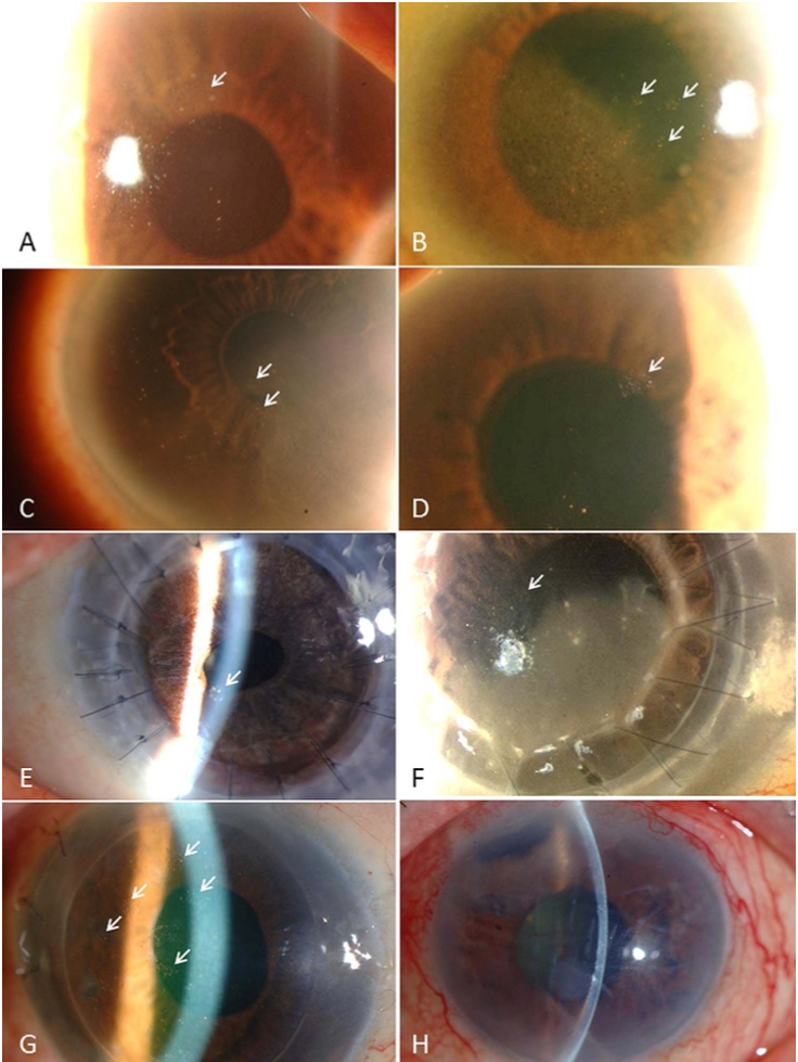
Representative anterior-segment photographs of cytomegalovirus corneal endotheliitis. (A-G) Typical CMV endotheliitis: CMV endotheliitis-associated coin-shaped lesions (white arrows). (H) Atypical CMV endotheliitis: CMV endotheliitis not associated with coin-shaped lesions. This case showed corneal oedema with keratic precipitates associated with recurrent anterior uveitis and secondary glaucoma.
Koizumi N, Inatomi T, Suzuki T for the Japan Corneal Endotheliitis Study Group, et al. Clinical features and management of cytomegalovirus corneal endotheliitis: analysis of 106 cases from the Japan corneal endotheliitis study. British Journal of Ophthalmology 2015;99:54-58.
File history
Click on a date/time to view the file as it appeared at that time.
| Date/Time | Thumbnail | Dimensions | User | Comment | |
|---|---|---|---|---|---|
| current | 12:38, March 16, 2021 | | 736 × 979 (1,013 KB) | Asim.farooq (talk | contribs) | Representative anterior-segment photographs of cytomegalovirus corneal endotheliitis. (A-G) Typical CMV endotheliitis: CMV endotheliitis-associated coin-shaped lesions (white arrows). (H) Atypical CMV endotheliitis: CMV endotheliitis not associated wit... |
You cannot overwrite this file.
File usage
The following page uses this file: