File:Increased FAZ nanopthalmos.png

Original file (1,136 × 914 pixels, file size: 969 KB, MIME type: image/png)
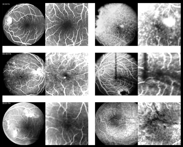
Fluorescein angiograms (FAs) of nanophthalmic patients, each of which shows a small, rudimentary foveal avascular zone (FAZ; image on right of each pair = 3x magnification of fovea). The top left images are from a normal 28-year-old female with a normal FAZ. Imaging of the left eye of nanophthalmic Patient 1 reveals a small, rudimentary FAZ and occult CNV (Top right). Nanophthalmic Patient 2’s FA shows a small, rudimentary FAZ, juxtafoveal window defect (fades late without leakage), and superior choroidal folds (2nd row left). Patient 2’s left eye has a small, rudimentary FAZ and choroidal folds (2nd row right, images). The FA from nanophthalmic Patient 3 shows a small, rudimentary FAZ and window defects OS (fade late without leakage; 3rd row left). Nanophthalmic Patient 4 also has a small, rudimentary FAZ OS (3rd row right, images). Ref: Walsh MK, Goldberg MF. Abnormal foveal avascular zone in nanophthalmos. Am J Ophthalmol. 2007 Jun;143(6):1067-1068.
File history
Click on a date/time to view the file as it appeared at that time.
| Date/Time | Thumbnail | Dimensions | User | Comment | |
|---|---|---|---|---|---|
| current | 08:38, December 6, 2023 | | 1,136 × 914 (969 KB) | Janine.Collinge (talk | contribs) |
You cannot overwrite this file.
File usage
The following page uses this file: