File:Image 3.png
From EyeWiki
No higher resolution available.
Image_3.png (530 × 211 pixels, file size: 164 KB, MIME type: image/png)
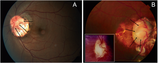
Fundus photographs demonstrating peripapillary staphyloma – A: Normal disc surrounded by peripapillary staphyloma. B: Close inspection reveals the optic disc is normal, as outlined by the arrows. The inset photograph shows a typical optic disc coloboma for comparison. Photo credit: Lingam G, Sen AC, Lingam V, Bhende M, Padhi TR, Xinyi S. Ocular coloboma-a comprehensive review for the clinician. Eye (Lond). 2021;35(8):2086-2109. doi:10.1038/s41433-021-01501-5; <a rel="license" href="http://creativecommons.org/licenses/by/4.0/"><img alt="Creative Commons License" style="border-width:0" src="https://i.creativecommons.org/l/by/4.0/80x15.png" /></a>
This work is licensed under a <a rel="license" href="http://creativecommons.org/licenses/by/4.0/">Creative Commons Attribution 4.0 International License</a>. No changes were made to this image.
File history
Click on a date/time to view the file as it appeared at that time.
| Date/Time | Thumbnail | Dimensions | User | Comment | |
|---|---|---|---|---|---|
| current | 09:17, June 20, 2022 | 530 × 211 (164 KB) | Mohammad.Pakravan (talk | contribs) |
You cannot overwrite this file.
File usage
The following page uses this file: