File:Image 2.png
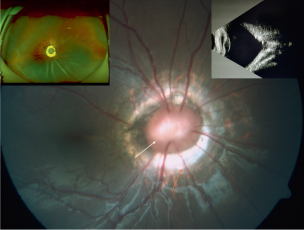
Image_2.png (304 × 230 pixels, file size: 101 KB, MIME type: image/png)
Image 2: Photographs of Morning Glory Disc - The white arrow points at the tuft of glial tissue at the center. The orange arrow at the bottom of the image points to the relatively straight blood vessels from the optic disc. The left inset photo shows retinal detachment in an eye with morning glory syndrome and the right inset photo is an ultrasound showing the funnel-shaped posterior excavation. Photo credit: Lingam G, Sen AC, Lingam V, Bhende M, Padhi TR, Xinyi S. Ocular coloboma-a comprehensive review for the clinician. Eye (Lond). 2021;35(8):2086-2109. doi:10.1038/s41433-021-01501-5; <a rel="license" href="http://creativecommons.org/licenses/by/4.0/"><img alt="Creative Commons License" style="border-width:0" src="https://i.creativecommons.org/l/by/4.0/80x15.png" /></a>
This work is licensed under a <a rel="license" href="http://creativecommons.org/licenses/by/4.0/">Creative Commons Attribution 4.0 International License</a>. No changes were made to this image.
File history
Click on a date/time to view the file as it appeared at that time.
| Date/Time | Thumbnail | Dimensions | User | Comment | |
|---|---|---|---|---|---|
| current | 09:00, June 20, 2022 | | 304 × 230 (101 KB) | Mohammad.Pakravan (talk | contribs) |
You cannot overwrite this file.
File usage
The following page uses this file: