File:IJO-64-415-g003.jpg
Original file (539 × 718 pixels, file size: 115 KB, MIME type: image/jpeg)
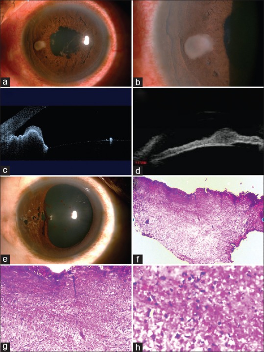
Figure 9 Excisional biopsy of iris tumor after thorough evaluation. [Digital images] by Rishi et al. Retrieved from https://www.ncbi.nlm.nih.gov/pmc/articles/PMC4991162/figure/F3/ Licensed under CC BY-NC-SA 3.0.
Original caption: “External photograph (a) of right eye shows circumcorneal congestion and greyish lesion in mid-peripheral iris of right eye. Slit-lamp photo (b) shows solid tumor of 2 mm × 1 mm size. Anterior segment optical coherence tomography (c) and ultrasound biomicroscopy (d) showed the tumor within iris stroma without extension to the posterior iris surface or angle. External photo (e) following excision biopsy of the tumor and primary repair of iris defect; pupil is spared. Histopathological examination (f-h) shows scattered lymphocytes and plasma cells without any granuloma formation (H and E, ×10, ×20, ×40)”
File history
Click on a date/time to view the file as it appeared at that time.
| Date/Time | Thumbnail | Dimensions | User | Comment | |
|---|---|---|---|---|---|
| current | 20:07, May 22, 2017 | | 539 × 718 (115 KB) | Frank.S.Hwang (talk | contribs) | Figure 9 Excisional biopsy of iris tumor after thorough evaluation. [Digital images] by Rishi et al. Retrieved from https://www.ncbi.nlm.nih.gov/pmc/articles/PMC4991162/figure/F3/ Licensed under CC BY-NC-SA 3.0. Original caption: “External photograp... |
You cannot overwrite this file.
File usage
The following page uses this file: