File:Hanif Figure 3 Multimodal.png

Original file (1,500 × 322 pixels, file size: 648 KB, MIME type: image/png)
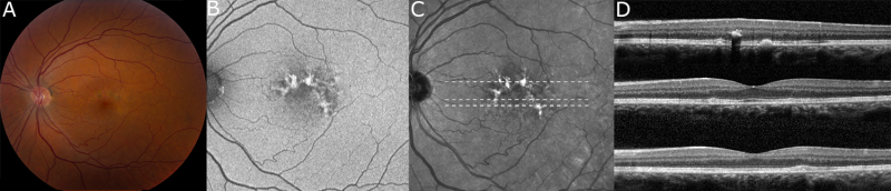
Multimodal imaging of macular pigment alterations in pentosan polysulfate maculopathy. Macular pigment clumps are visible in color fundus photography (A) and colocalize with hyperautofluorescent spots on fundus autofluorescence imaging (B) and hyperreflectant spots on near-infrared reflectance imaging (C). The lesions cast a shadow on the underlying choroid on ocular coherence tomography (D) and appear to be located in the retinal pigment epithelium. The locations of the scans in panel D correspond to the dotted lines in panel C in descending order.
File history
Click on a date/time to view the file as it appeared at that time.
| Date/Time | Thumbnail | Dimensions | User | Comment | |
|---|---|---|---|---|---|
| current | 13:20, January 22, 2021 | 1,500 × 322 (648 KB) | Nieraj.Jain (talk | contribs) | Multimodal imaging of macular pigment alterations in pentosan polysulfate maculopathy. Macular pigment clumps are visible in color fundus photography (A) and colocalize with hyperautofluorescent spots on fundus autofluorescence imaging (B) and hyperref... | |
| 07:05, January 22, 2021 | 1,500 × 322 (648 KB) | Nieraj.Jain (talk | contribs) | |||
| 06:57, January 22, 2021 | 1,956 × 486 (1.08 MB) | Nieraj.Jain (talk | contribs) | Multimodal imaging of macular pigment alterations in pentosan polysulfate maculopathy. Macular pigment clumps are visible in color fundus photography (A) and colocalize with hyperautofluorescent spots on fundus autofluorescence imaging (B) and hyperref... |
You cannot overwrite this file.
File usage
The following page uses this file: