File:Figure 8 AS OCT.jpeg
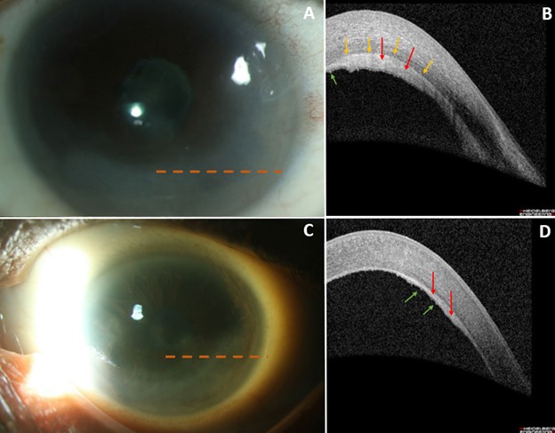
Figure_8_AS_OCT.jpeg (624 × 487 pixels, file size: 56 KB, MIME type: image/jpeg)
Summary
Figures 8A and 8C: Demonstrate slit lamp photos of the right and left eye, respectively. There is inferior corneal opacification (haze) and the dashed orange lines represent the cross-section of the corresponding AS-OCT images. Figure 8B: AS-OCT of the right eye shows a plaque involving the endothelium and deep stroma (red arrows) with a prominent hyperreflective line between the plaque and deep stroma (yellow arrows), which is more distinct than Descemet’s membrane. Figure 8D: OCT of the OS showing a similar albeit less thickened endothelial-stromal plaque with associated fine hyperreflective keratic precipitates (green arrows).
File history
Click on a date/time to view the file as it appeared at that time.
| Date/Time | Thumbnail | Dimensions | User | Comment | |
|---|---|---|---|---|---|
| current | 18:14, October 18, 2022 | | 624 × 487 (56 KB) | Justin.Yamanuha (talk | contribs) | Figures 8A and 8C: Demonstrate slit lamp photos of the right and left eye, respectively. There is inferior corneal opacification (haze) and the dashed orange lines represent the cross-section of the corresponding AS-OCT images. Figure 8B: AS-OCT of the right eye shows a plaque involving the endothelium and deep stroma (red arrows) with a prominent hyperreflective line between the plaque and deep stroma (yellow arrows), which is more distinct than Descemet’s membrane. Figure 8D: OCT of the OS... |
You cannot overwrite this file.
File usage
The following page uses this file: