File:Figure 6 Slit Lamp Photo HSV Dendrite.jpg
From EyeWiki

No higher resolution available.
Figure_6_Slit_Lamp_Photo_HSV_Dendrite.jpg (713 × 469 pixels, file size: 42 KB, MIME type: image/jpeg)
Figure 6. Herpes Keratitis. Under cobalt-blue illumination, fluorescein bound to the basement membrane underlying damaged corneal epithelium fluoresces bright green. This slit lamp photograph illustrates the unique “dendritic” branching lesions characteristic of herpes keratitis.
File history
Click on a date/time to view the file as it appeared at that time.
| Date/Time | Thumbnail | Dimensions | User | Comment | |
|---|---|---|---|---|---|
| current | 13:19, May 28, 2020 | | 713 × 469 (42 KB) | Justin.Yamanuha (talk | contribs) | Reverted to version as of 20:11, May 28, 2020 |
| 13:15, May 28, 2020 |  | 713 × 469 (42 KB) | Justin.Yamanuha (talk | contribs) | Figure 6. Herpes Keratitis. Under cobalt-blue illumination, fluorescein bound to the basement membrane underlying damaged corneal epithelium fluoresces bright green. This slit lamp photograph illustrates the unique “dendritic” branching lesions ch... | |
| 13:11, May 28, 2020 | 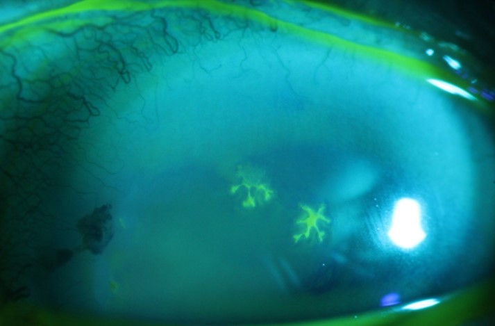 | 713 × 469 (42 KB) | Justin.Yamanuha (talk | contribs) | Figure 6. Slit lamp modified with a large plexiglass breath shield to provide a barrier to respiratory transmission. Numbers are referenced in the text description. |
You cannot overwrite this file.
File usage
The following page uses this file: