File:Figure 6 Eyewiki.png

Original file (1,080 × 808 pixels, file size: 1.49 MB, MIME type: image/png)
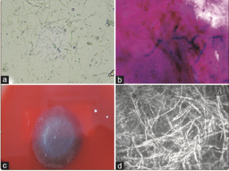
(a) Shows a 10% KOH wet mount demonstrating the presence of long, sparsely septate hyaline hyphae. (b) Shows the gram stain image depicting the thick cell wall, a few septate, and ribbon-like folding patterns of fungal hyphae. (c) Shows a 5-day old culture of P. insidiosum at 37°C grown on 5% sheep blood agar. (d) Shows a confocal microscopy image depicting thin, hyperreflective, occasionally branching structures with varying angles
Reproduced from permission by Editor in Chief- IJO (Dr.SH)
Source- Gurnani B, Christy J, Narayana S, Rajkumar P, Kaur K, Gubert J. Retrospective multifactorial analysis of Pythium keratitis and review of literature. Indian J Ophthalmol. 2021 May;69(5):1095-1101. doi: 10.4103/ijo.IJO_1808_20. PMID: 33913840; PMCID: PMC8186601.
File history
Click on a date/time to view the file as it appeared at that time.
| Date/Time | Thumbnail | Dimensions | User | Comment | |
|---|---|---|---|---|---|
| current | 23:21, May 27, 2023 | | 1,080 × 808 (1.49 MB) | Bharat.Gurnani (talk | contribs) |
You cannot overwrite this file.
File usage
The following page uses this file: