File:Figure 6 AS OCT.jpeg
From EyeWiki
No higher resolution available.
Figure_6_AS_OCT.jpeg (624 × 229 pixels, file size: 35 KB, MIME type: image/jpeg)
Summary
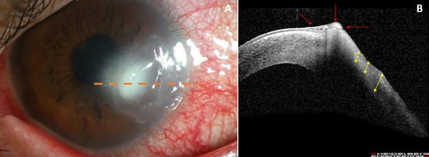
Figure 6A: Slit lamp photo of the left eye showing a new infiltrate adjacent to an area of prior corneal thinning in a patient with a neurotrophic cornea. The orange dotted line in 6A represents the plane of 6B. Figure 6B: AS-OCT image of the left eye demonstrating heaped hyperreflective epithelium (red arrows) adjacent to an area of irregular thin stroma from a prior episode of infectious keratitis (yellow arrows).
File history
Click on a date/time to view the file as it appeared at that time.
| Date/Time | Thumbnail | Dimensions | User | Comment | |
|---|---|---|---|---|---|
| current | 18:05, October 18, 2022 | 624 × 229 (35 KB) | Justin.Yamanuha (talk | contribs) | Figure 6A: Slit lamp photo of the left eye showing a new infiltrate adjacent to an area of prior corneal thinning in a patient with a neurotrophic cornea. The orange dotted line in 6A represents the plane of 6B. Figure 6B: AS-OCT image of the left eye demonstrating heaped hyperreflective epithelium (red arrows) adjacent to an area of irregular thin stroma from a prior episode of infectious keratitis (yellow arrows). |
You cannot overwrite this file.
File usage
The following page uses this file: