File:Figure 5 new.png
Original file (1,070 × 708 pixels, file size: 1.28 MB, MIME type: image/png)
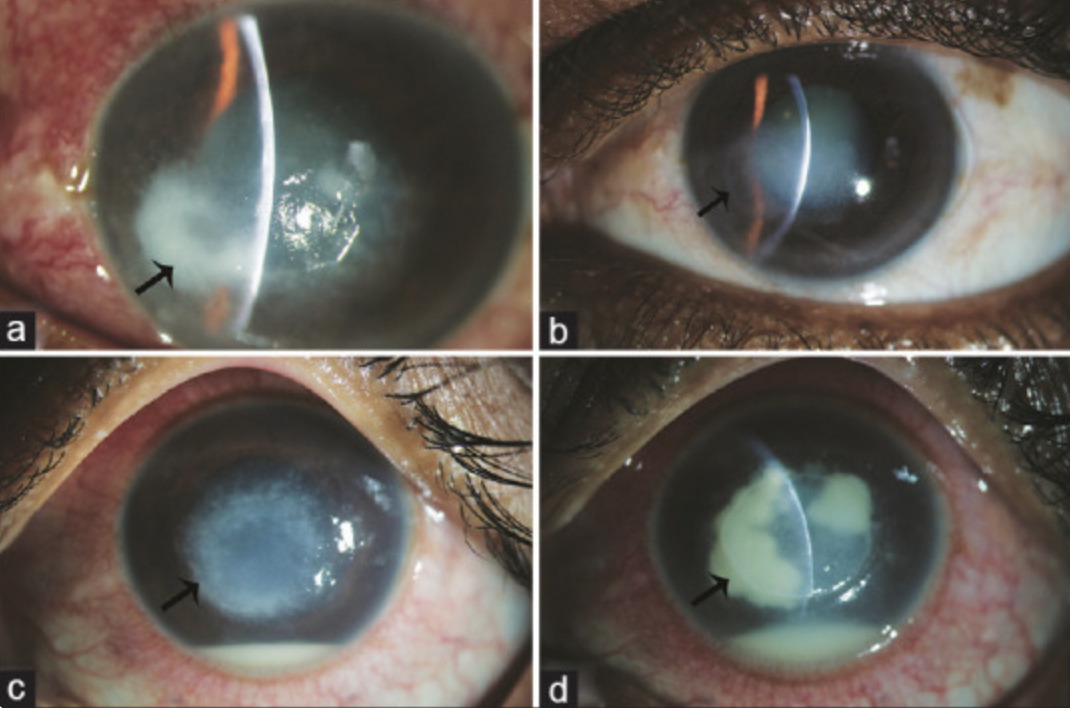
(a) Slit-lamp image depicting the case of confirmed Pythium keratitis having anterior to mid stromal infiltrate with tentacular extensions. (b) Slit-lamp image depicting the image of the same case after resolution on medical treatment. (c) Slit-lamp image depicting the case of confirmed Pythium keratitis having anterior to mid stromal infiltrate having tentacular extensions extending till posterior stroma. (d) Slit-lamp image depicting worsening of infection as observed by increased density of endo-exudates with cotton wool-like fluffy infiltrates
Reproduced with permission from Editor in Chief -IJO (Dr. SH)
Source-Gurnani B, Kaur K, Venugopal A, Srinivasan B, Bagga B, Iyer G, Christy J, Prajna L, Vanathi M, Garg P, Narayana S, Agarwal S, Sahu S. Pythium insidiosum keratitis - A review. Indian J Ophthalmol. 2022 Apr;70(4):1107-1120. doi: 10.4103/ijo.IJO_1534_21. PMID: 35325996; PMCID: PMC9240499.
File history
Click on a date/time to view the file as it appeared at that time.
| Date/Time | Thumbnail | Dimensions | User | Comment | |
|---|---|---|---|---|---|
| current | 23:37, May 27, 2023 | | 1,070 × 708 (1.28 MB) | Bharat.Gurnani (talk | contribs) |
You cannot overwrite this file.
File usage
The following page uses this file: