File:Figure 5 SWM.jpg
From EyeWiki
No higher resolution available.
Figure_5_SWM.jpg (435 × 458 pixels, file size: 44 KB, MIME type: image/jpeg)
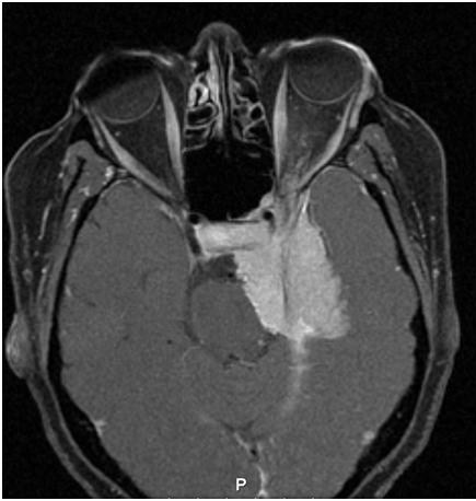
FIGURE 5: Axial T1 post gadolinium MRI image showing a diffuse sphenoid wing meningioma involving the left orbital apex, and cavernous sinus exerting a mass effect on the pons and orbital structures. The image shows the typical appearance of meningioma signified by thickening with sparing of adjacent anatomical structures, as well as exhibiting prominent contrast enhancement and a dural tail.
File history
Click on a date/time to view the file as it appeared at that time.
| Date/Time | Thumbnail | Dimensions | User | Comment | |
|---|---|---|---|---|---|
| current | 17:03, November 2, 2013 | | 435 × 458 (44 KB) | Kenneth.M.Downes (talk | contribs) | |
| 16:33, November 2, 2013 | 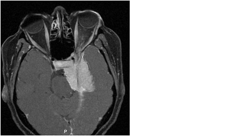 | 800 × 458 (29 KB) | Kenneth.M.Downes (talk | contribs) | FIGURE 5: Axial T1 post gadolinium MRI image showing a diffuse sphenoid wing meningioma involving the left orbital apex, and cavernous sinus exerting a mass effect on the pons and orbital structures. The image shows the typical appearance of meningioma s |
You cannot overwrite this file.
File usage
The following page uses this file: