File:Figure 4 Eyewiki.png

Original file (1,620 × 1,268 pixels, file size: 3.45 MB, MIME type: image/png)
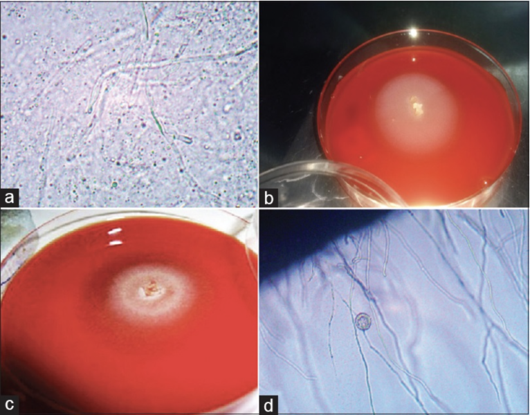
(a)10% KOH wet mount preparation of corneal scraping of Pythium insidiosum showing linear elongated sparsely septate hyaline hyphae. A number of small vesicles within the hyphae are also observed (b) A 3-day old subculture of P. insidiosum at 37°C grown on 5% sheep blood agar (c) A 5-day old subculture of P. insidiosum at 37°C grown on 5% sheep blood agar[29] (d) A small round vesicle with numerous zoospores that developed after 3 h of incubation before zoospore release (×10) using cultured leaf incarnation method
Reproduced by permission from Editor in Chief IJO (Dr. SH)
Source-Gurnani B, Christy J, Narayana S, Rajkumar P, Kaur K, Gubert J. Retrospective multifactorial analysis of Pythium keratitis and review of literature. Indian J Ophthalmol. 2021 May;69(5):1095-1101. doi: 10.4103/ijo.IJO_1808_20. PMID: 33913840; PMCID: PMC8186601.
File history
Click on a date/time to view the file as it appeared at that time.
| Date/Time | Thumbnail | Dimensions | User | Comment | |
|---|---|---|---|---|---|
| current | 23:01, May 27, 2023 | | 1,620 × 1,268 (3.45 MB) | Bharat.Gurnani (talk | contribs) |
You cannot overwrite this file.
File usage
The following page uses this file: