File:Figure 3 Slit Lamp Photo Episcleritis.jpg
From EyeWiki
No higher resolution available.
Figure_3_Slit_Lamp_Photo_Episcleritis.jpg (663 × 533 pixels, file size: 66 KB, MIME type: image/jpeg)
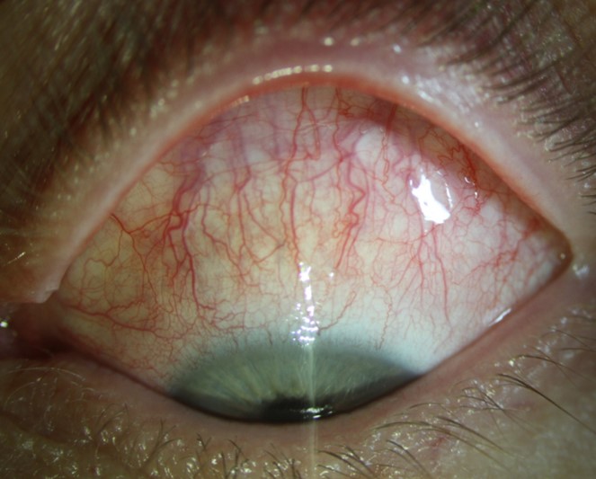
Figure 3. Diffuse Episcleritis. Slit lamp photograph under diffuse illumination shows injection due to engorged episcleral vessels that course radially beneath the conjunctiva toward the limbus. Pain is typically absent or mild compared with scleritis.
File history
Click on a date/time to view the file as it appeared at that time.
| Date/Time | Thumbnail | Dimensions | User | Comment | |
|---|---|---|---|---|---|
| current | 13:09, May 28, 2020 | | 663 × 533 (66 KB) | Justin.Yamanuha (talk | contribs) | Figure 3. Diffuse Episcleritis. Slit lamp photograph under diffuse illumination shows injection due to engorged episcleral vessels that course radially beneath the conjunctiva toward the limbus. Pain is typically absent or mild compared with scleritis. |
You cannot overwrite this file.
File usage
The following page uses this file: