File:Figure 3 Eyewiki.png
Original file (1,654 × 956 pixels, file size: 2.09 MB, MIME type: image/png)
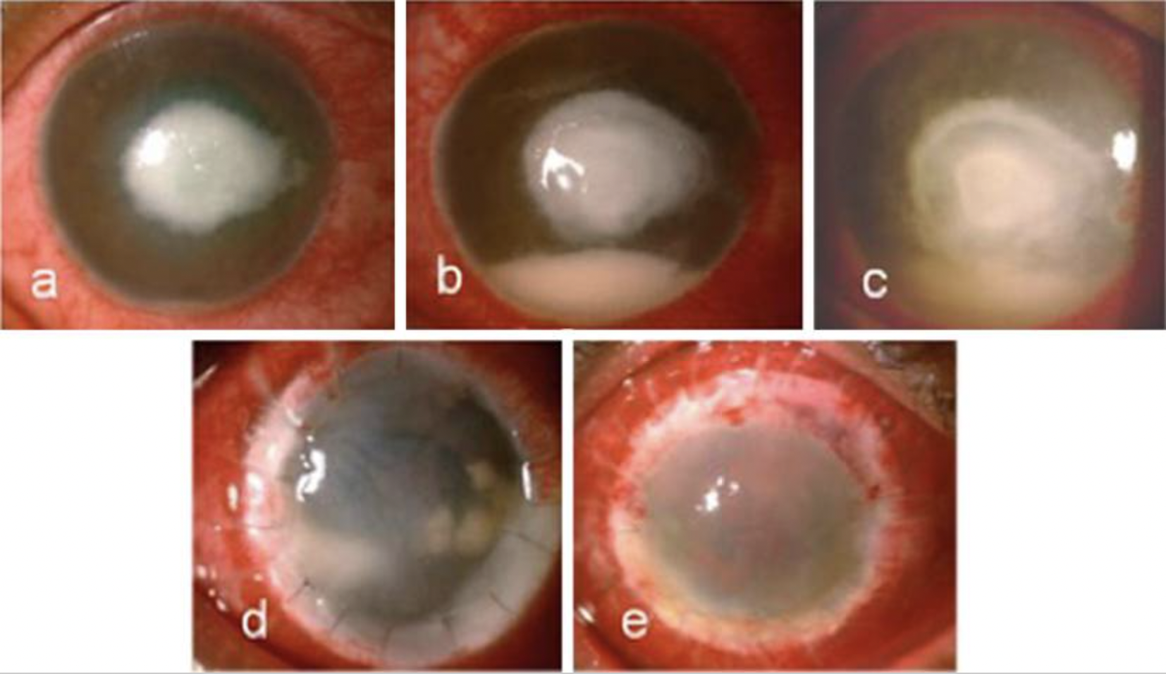
Digital image of a patient with rapidly proliferative Pythium insidiosum keratitis. a At presentation (day 1)—5 × 6 mm central full-thickness infiltrate with trace hypopyon. b, c (day 7) Worsening of full-thickness infiltrate with rapid spread towards limbus and increase size and density of hypopyon despite topical medications. d Recurrence-graft infection noted 7 days following therapeutic penetrating keratoplasty, e 1 month following a regraft-diffuse congestion, stromal edema, and 360-degree superficial vascularization
Reproduced by permission from Editor-in-Chief- Ophthalmology and Therapy
Source-Gurnani B, Kaur K, Agarwal S, Lalgudi VG, Shekhawat NS, Venugopal A, Tripathy K, Srinivasan B, Iyer G, Gubert J. Pythium insidiosum Keratitis: Past, Present, and Future. Ophthalmol Ther. 2022 Oct;11(5):1629-1653. doi: 10.1007/s40123-022-00542-7. Epub 2022 Jul 5. PMID: 35788551; PMCID: PMC9255487.
File history
Click on a date/time to view the file as it appeared at that time.
| Date/Time | Thumbnail | Dimensions | User | Comment | |
|---|---|---|---|---|---|
| current | 22:50, May 27, 2023 | | 1,654 × 956 (2.09 MB) | Bharat.Gurnani (talk | contribs) |
You cannot overwrite this file.
File usage
The following page uses this file: