File:Figure 3B.jpg
From EyeWiki
Size of this preview: 700 × 599 pixels. Other resolution: 2,392 × 2,048 pixels.
Original file (2,392 × 2,048 pixels, file size: 816 KB, MIME type: image/jpeg)
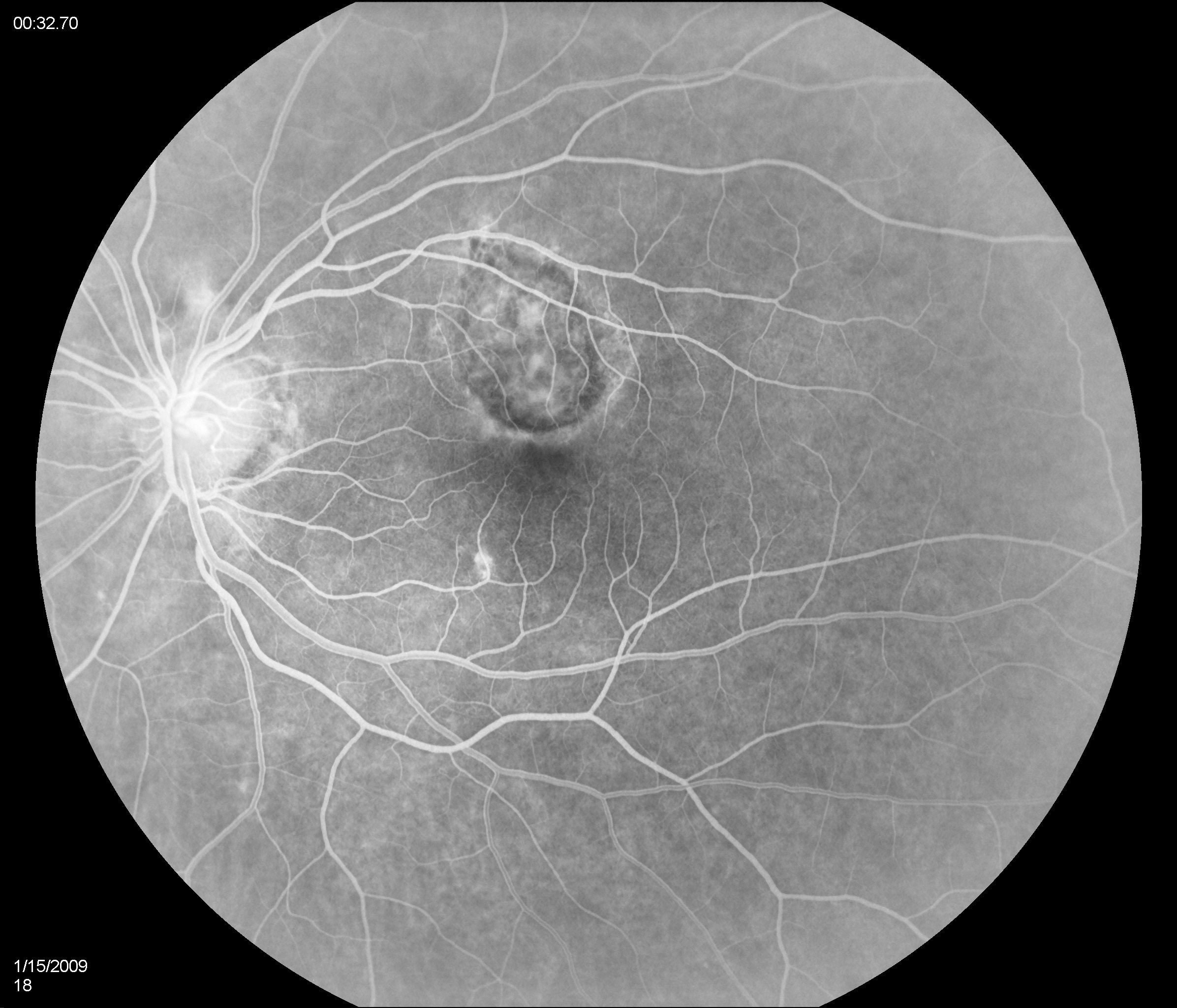
Early phase of the fluorescein angiogram reveals hyperfluorescence corresponding to the CNV lesion with hypofluorescent borders corresponding to the pigmented borders of the lesion.
Courtesy of Dr Narsing Rao and taken from the following publication: Pachydaki SI, Jakobiec FA, Bhat P, Sobrin L, Michaud NA, Seshan SV, D'Amico DJ. Surgical management and ultrastructural study of choroidal neovascularization in punctate inner choroidopathy after bevacizumab. J Ophthalmic Inflamm Infect. 2011 Nov 27.
File history
Click on a date/time to view the file as it appeared at that time.
| Date/Time | Thumbnail | Dimensions | User | Comment | |
|---|---|---|---|---|---|
| current | 21:15, November 30, 2011 | | 2,392 × 2,048 (816 KB) | Katrina.A.Mears (talk | contribs) | Early phase of the fluorescein angiogram reveals hyperfluorescence corresponding to the CNV lesion with hypofluorescent borders corresponding to the pigmented borders of the lesion. Courtesy of Dr Narsing Rao and taken from the following publication: |
You cannot overwrite this file.
File usage
The following page uses this file: