File:Figure 2 AS OCT.jpeg
From EyeWiki
No higher resolution available.
Figure_2_AS_OCT.jpeg (624 × 167 pixels, file size: 29 KB, MIME type: image/jpeg)
Summary
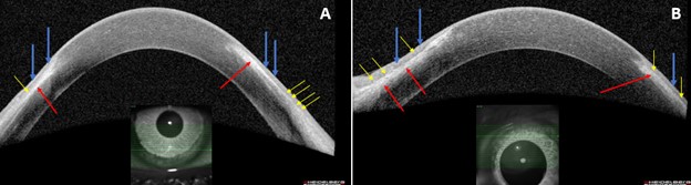
Figure 2A and 2B: AS-OCT of the right eye (left image) and AS-OCT of the left eye (right image) demonstrate healed PUK adjacent to the limbus in both eyes. Images show thinned hyporeflective epithelium (blue arrows) overlying focal areas of stromal hyperreflectivity (red arrows) with a sharp line of demarcation between the two regions (yellow arrows).
File history
Click on a date/time to view the file as it appeared at that time.
| Date/Time | Thumbnail | Dimensions | User | Comment | |
|---|---|---|---|---|---|
| current | 17:46, October 18, 2022 | 624 × 167 (29 KB) | Justin.Yamanuha (talk | contribs) | Figure 2A and 2B: AS-OCT of the right eye (left image) and AS-OCT of the left eye (right image) demonstrate healed PUK adjacent to the limbus in both eyes. Images show thinned hyporeflective epithelium (blue arrows) overlying focal areas of stromal hyperreflectivity (red arrows) with a sharp line of demarcation between the two regions (yellow arrows). |
You cannot overwrite this file.
File usage
The following page uses this file: