File:Figure 2.png
From EyeWiki
Figure_2.png (315 × 442 pixels, file size: 102 KB, MIME type: image/png)
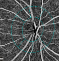
Figure 2 - Normal optic disc microvasculature imaged using OCT-A.
File history
Click on a date/time to view the file as it appeared at that time.
| Date/Time | Thumbnail | Dimensions | User | Comment | |
|---|---|---|---|---|---|
| current | 15:29, January 14, 2018 |  | 315 × 442 (102 KB) | Aroucha.Vickers (talk | contribs) | |
| 02:50, June 2, 2017 |  | 979 × 1,000 (1.06 MB) | David.Samuel.Cordeiro.Sousa (talk | contribs) | Reverted to version as of 00:11, May 22, 2017 | |
| 21:06, June 1, 2017 |  | 1,937 × 995 (2.92 MB) | M.Margarita.Parra (talk | contribs) | A. Color image of fovea, melanocytoma is partially visible. B and C. En face OCT and OCT angiograms of inner retinal plexus, revealed a honeycomb pattern (green area) corresponding to intraretinal cysts and hyper-reflective dots (blue area) around the... | |
| 16:11, May 21, 2017 |  | 979 × 1,000 (1.06 MB) | David.Samuel.Cordeiro.Sousa (talk | contribs) | Figure 2 - Normal optic disc microvasculature imaged using OCT-A. |
You cannot overwrite this file.
File usage
The following page uses this file:


