File:Figure 10 AS OCT.jpeg
From EyeWiki
No higher resolution available.
Figure_10_AS_OCT.jpeg (624 × 368 pixels, file size: 45 KB, MIME type: image/jpeg)
Summary
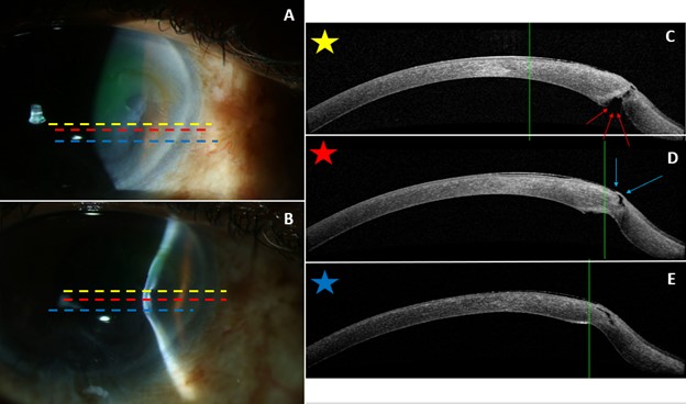
Figure 10A and 10B demonstrate slit lamp photos of the right eye. The dashed lines represent the cross-sections of the AS-OCT images (10C, 10D, 10E). The dashed line colors correspond to star colors. The red arrows in 10C show a focal path from the deep stroma anteriorly with only a thin area of overlying intact epithelium (blue arrows in 10D).
File history
Click on a date/time to view the file as it appeared at that time.
| Date/Time | Thumbnail | Dimensions | User | Comment | |
|---|---|---|---|---|---|
| current | 18:21, October 18, 2022 | | 624 × 368 (45 KB) | Justin.Yamanuha (talk | contribs) | Figure 10A and 10B demonstrate slit lamp photos of the right eye. The dashed lines represent the cross-sections of the AS-OCT images (10C, 10D, 10E). The dashed line colors correspond to star colors. The red arrows in 10C show a focal path from the deep stroma anteriorly with only a thin area of overlying intact epithelium (blue arrows in 10D). |
You cannot overwrite this file.
File usage
The following page uses this file: