File:Figure 1..png
From EyeWiki
No higher resolution available.
Figure_1..png (523 × 234 pixels, file size: 221 KB, MIME type: image/png)
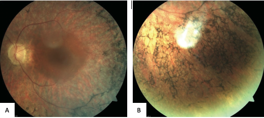
Figure 1. Fundus photograph of the left eye with UPR [8] A. Fundus photograph of left eye B. Fundus photograph of the periphery of the left eye Waxy disc pallor, markedly attenuated retinal arterioles and clumps of bone-spicule pigments scattered in the mid periphery, in all the quadrants of the retina
File history
Click on a date/time to view the file as it appeared at that time.
| Date/Time | Thumbnail | Dimensions | User | Comment | |
|---|---|---|---|---|---|
| current | 18:16, September 13, 2022 | | 523 × 234 (221 KB) | MarieH.Errera (talk | contribs) |
You cannot overwrite this file.
File usage
The following page uses this file: