File:Fig 2 Eyewiki.png
Original file (1,616 × 1,288 pixels, file size: 3.28 MB, MIME type: image/png)
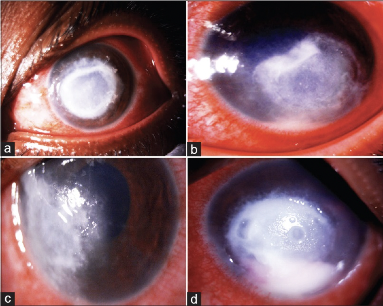
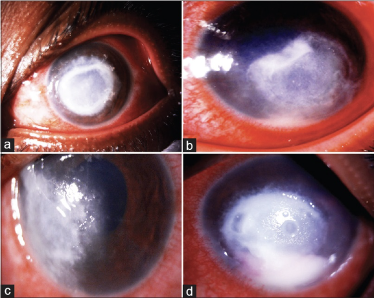
(a) Slit-lamp image of the cornea showing central, dense, greyish-white circular 8x8x mm infiltrate with peripheral tentacular projections (b) Image depicting diffuse white stromal infiltrates. In addition peripheral furrowing is seen in this image inferiorly from 4 o clock to 6 o clock hour (c) Image depicting diffuse white stromal infiltrates showing a tendency for limbal spread (d) Image depicting diffuse white full thickness infiltrates along with anterior chamber hypopyon
Reproduced with permission from the Editor in Chief IJO (Dr SH)
Source-Gurnani B, Christy J, Narayana S, Rajkumar P, Kaur K, Gubert J. Retrospective multifactorial analysis of Pythium keratitis and review of literature. Indian J Ophthalmol. 2021 May;69(5):1095-1101. doi: 10.4103/ijo.IJO_1808_20. PMID: 33913840; PMCID: PMC8186601.
File history
Click on a date/time to view the file as it appeared at that time.
| Date/Time | Thumbnail | Dimensions | User | Comment | |
|---|---|---|---|---|---|
| current | 22:45, May 27, 2023 | | 1,616 × 1,288 (3.28 MB) | Bharat.Gurnani (talk | contribs) |
You cannot overwrite this file.
File usage
The following page uses this file: