File:Fig3.jpg
From EyeWiki
No higher resolution available.
Fig3.jpg (624 × 351 pixels, file size: 44 KB, MIME type: image/jpeg)
Summary
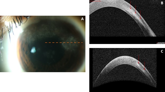
Figure 3A: Slit lamp photo of the left eye at presentation with non-infectious interstitial keratitis showing focal areas of corneal haze. Dashed orange line is the plane of both AS-OCTs in 3B and 3C. Figure 3B: AS-OCT at presentation with focal areas of discrete mid-stromal hyperreflectivity with associated overlying and underlying stromal hyporeflectivity. Figure 3C: AS-OCT demonstrating more compact areas of mid-stromal hyperreflectivity (red arrows) after treatment with topical prednisolone and systemic methotrexate.
File history
Click on a date/time to view the file as it appeared at that time.
| Date/Time | Thumbnail | Dimensions | User | Comment | |
|---|---|---|---|---|---|
| current | 17:50, October 18, 2022 | | 624 × 351 (44 KB) | Justin.Yamanuha (talk | contribs) | Figure 3A: Slit lamp photo of the left eye at presentation with non-infectious interstitial keratitis showing focal areas of corneal haze. Dashed orange line is the plane of both AS-OCTs in 3B and 3C. Figure 3B: AS-OCT at presentation with focal areas of discrete mid-stromal hyperreflectivity with associated overlying and underlying stromal hyporeflectivity. Figure 3C: AS-OCT demonstrating more compact areas of mid-stromal hyperreflectivity (red arrows) after treatment with topical prednisolo... |
You cannot overwrite this file.
File usage
The following page uses this file: