File:FLIGHT2.jpg
FLIGHT2.jpg (432 × 238 pixels, file size: 33 KB, MIME type: image/jpeg)
Summary
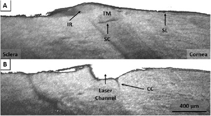
(A) SD-OCT showing the intact TM and Schlemm's canal (SC) in the region adjacent to the FLT drainage channel. Schwalbe's line (SL) and residual iris root (IR) are also visible. (B) SD-OCT shows the FLT channel penetrating Schlemm's canal through the trabecular meshwork. Also visible is a large collector channel within the region of the FLT channel. Reprinted with permission from: Mikula ER, Raksi F, Ahmed II, et al. Femtosecond Laser Trabeculotomy in Perfused Human Cadaver Anterior Segments: A Novel, Noninvasive Approach to Glaucoma Treatment. Transl Vis Sci Technol. 2022;11(3):28.
File history
Click on a date/time to view the file as it appeared at that time.
| Date/Time | Thumbnail | Dimensions | User | Comment | |
|---|---|---|---|---|---|
| current | 10:23, June 5, 2023 | | 432 × 238 (33 KB) | Abdelrahman.Elhusseiny (talk | contribs) | (A) SD-OCT showing the intact TM and Schlemm's canal (SC) in the region adjacent to the FLT drainage channel. Schwalbe's line (SL) and residual iris root (IR) are also visible. (B) SD-OCT shows the FLT channel penetrating Schlemm's canal through the trabecular meshwork. Also visible is a large collector channel within the region of the FLT channel. Reprinted with permission from: Mikula ER, Raksi F, Ahmed II, et al. Femtosecond Laser Trabeculotomy in Perfused Human Cadaver Anterior Segments:... |
You cannot overwrite this file.
File usage
The following page uses this file: