File:FLIGHT1.jpg
FLIGHT1.jpg (709 × 573 pixels, file size: 78 KB, MIME type: image/jpeg)
Summary
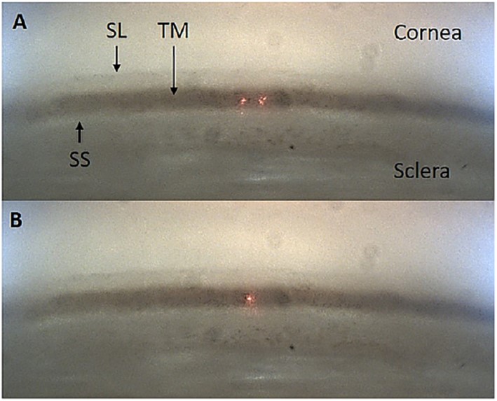
A typical ViaLase Inc. gonioscopic image acquired through an intact ex vivo perfused human anterior segment. The cornea, sclera, Schwalbe's line (SL), trabecular meshwork (TM), and scleral spur (SS) are clearly visible. The iris has been excised. (A) The dual aiming beams on the TM. (B) The dual aiming beams overlapped to a single point on the TM, indicating the focal plane of the femtosecond laser. Reprinted with permission from: Mikula ER, Raksi F, Ahmed II, et al. Femtosecond Laser Trabeculotomy in Perfused Human Cadaver Anterior Segments: A Novel, Noninvasive Approach to Glaucoma Treatment. Transl Vis Sci Technol. 2022;11(3):28.
File history
Click on a date/time to view the file as it appeared at that time.
| Date/Time | Thumbnail | Dimensions | User | Comment | |
|---|---|---|---|---|---|
| current | 10:21, June 5, 2023 | | 709 × 573 (78 KB) | Abdelrahman.Elhusseiny (talk | contribs) | A typical ViaLase Inc. gonioscopic image acquired through an intact ex vivo perfused human anterior segment. The cornea, sclera, Schwalbe's line (SL), trabecular meshwork (TM), and scleral spur (SS) are clearly visible. The iris has been excised. (A) The dual aiming beams on the TM. (B) The dual aiming beams overlapped to a single point on the TM, indicating the focal plane of the femtosecond laser. Reprinted with permission from: Mikula ER, Raksi F, Ahmed II, et al. Femtosecond Laser Trabecu... |
You cannot overwrite this file.
File usage
The following page uses this file: