File:FIG 1 REV.jpg
From EyeWiki

Size of this preview: 800 × 294 pixels. Other resolution: 1,873 × 688 pixels.
Original file (1,873 × 688 pixels, file size: 127 KB, MIME type: image/jpeg)
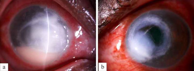
Figure 7 a- Digital slit lamp image of the right eye of the patient depicting diffuse conjunctival congestion, 8x8 mm creamy white full thickness infiltrate, stromal melt, nasal peripheral furrowing with guttering along with 3 mm anterior chamber hypopyon. b- Digital slit lamp image of the right eye of the patient depicting diffuse conjunctival congestion, crescentic eccentric 7x7 mm mid to posterior stromal infiltrate, inferonasal full thickness infiltrate, nasal limbal spread, central corneal thinning with impending perforation
File history
Click on a date/time to view the file as it appeared at that time.
| Date/Time | Thumbnail | Dimensions | User | Comment | |
|---|---|---|---|---|---|
| current | 03:26, May 30, 2023 | 1,873 × 688 (127 KB) | Bharat.Gurnani (talk | contribs) |
You cannot overwrite this file.
File usage
The following page uses this file: